
Lesson no 1 : Examine biochemical mechanisms of metabolic diseases such as diabetes and obesity.
The lesson, Examine biochemical mechanisms of metabolic diseases such as diabetes and obesity, provides Learners with an in-depth understanding of the complex biochemical processes that contribute to the development, progression and clinical consequences of major metabolic disorders. Metabolic diseases arise when normal pathways responsible for regulating energy production, glucose utilisation, lipid metabolism and hormonal signalling become disrupted. Understanding these mechanisms is essential for interpreting disease processes and supporting evidence-based approaches to prevention and therapeutic management.
A central focus of this lesson is the biochemical basis of diabetes, including the regulation of blood glucose, insulin secretion, insulin sensitivity and cellular glucose uptake. Learners will explore how impaired insulin action and pancreatic dysfunction can lead to persistent hyperglycaemia and associated metabolic complications. The lesson also examines obesity as a complex metabolic condition involving long-term energy imbalance, altered lipid storage, adipose tissue dysfunction, inflammation and changes in hormonal regulation.
The relationship between obesity and insulin resistance will be explored in detail, highlighting how excessive adipose tissue can influence inflammatory pathways, lipid metabolism and cellular signalling. Learners will also consider the roles of key organs, including the pancreas, liver, skeletal muscle and adipose tissue, in maintaining metabolic homeostasis. When communication between these systems becomes impaired, significant disturbances may develop that increase the risk of chronic metabolic disease.
In addition, the lesson introduces important biochemical concepts such as glucose homeostasis, lipolysis, lipogenesis, oxidative stress, mitochondrial function and inflammatory signalling. Learners will develop the ability to connect molecular and cellular mechanisms with observable clinical features and laboratory findings.
By critically examining the biochemical mechanisms underlying diabetes and obesity, Learners will gain a stronger foundation for understanding metabolic disease assessment, prevention and therapeutic strategies. This knowledge is particularly valuable for professionals working in clinical nutrition, healthcare, biomedical science and related disciplines, where the ability to interpret metabolic dysfunction supports safe, evidence-informed and person-centred practice.
1.Critically Examine the Progressive Biochemical Disruptions in Glucose and Lipid Metabolism That Characterise the Clinical Onset of Type 2 Diabetes
Type 2 diabetes mellitus is a complex and progressive metabolic disorder characterised primarily by persistent disturbances in glucose regulation. However, the clinical onset of the condition cannot be understood by examining glucose metabolism alone. Significant alterations in lipid metabolism, insulin signalling, hepatic function, adipose tissue activity and pancreatic beta-cell function occur gradually and interact with one another over time.
The progression towards type 2 diabetes commonly begins years before the condition is clinically diagnosed. During the early stages, the body may compensate for reduced insulin sensitivity by increasing insulin secretion. As metabolic dysfunction progresses, this compensatory mechanism becomes insufficient. Glucose concentrations begin to rise, lipid abnormalities become more pronounced, and pancreatic beta cells may progressively lose their capacity to maintain normal glucose homeostasis.
A critical understanding of these biochemical disruptions enables Learners to connect molecular mechanisms with clinical findings, laboratory markers and therapeutic approaches. It also demonstrates why type 2 diabetes should be viewed as a multi-system metabolic disorder rather than simply a condition of elevated blood glucose.
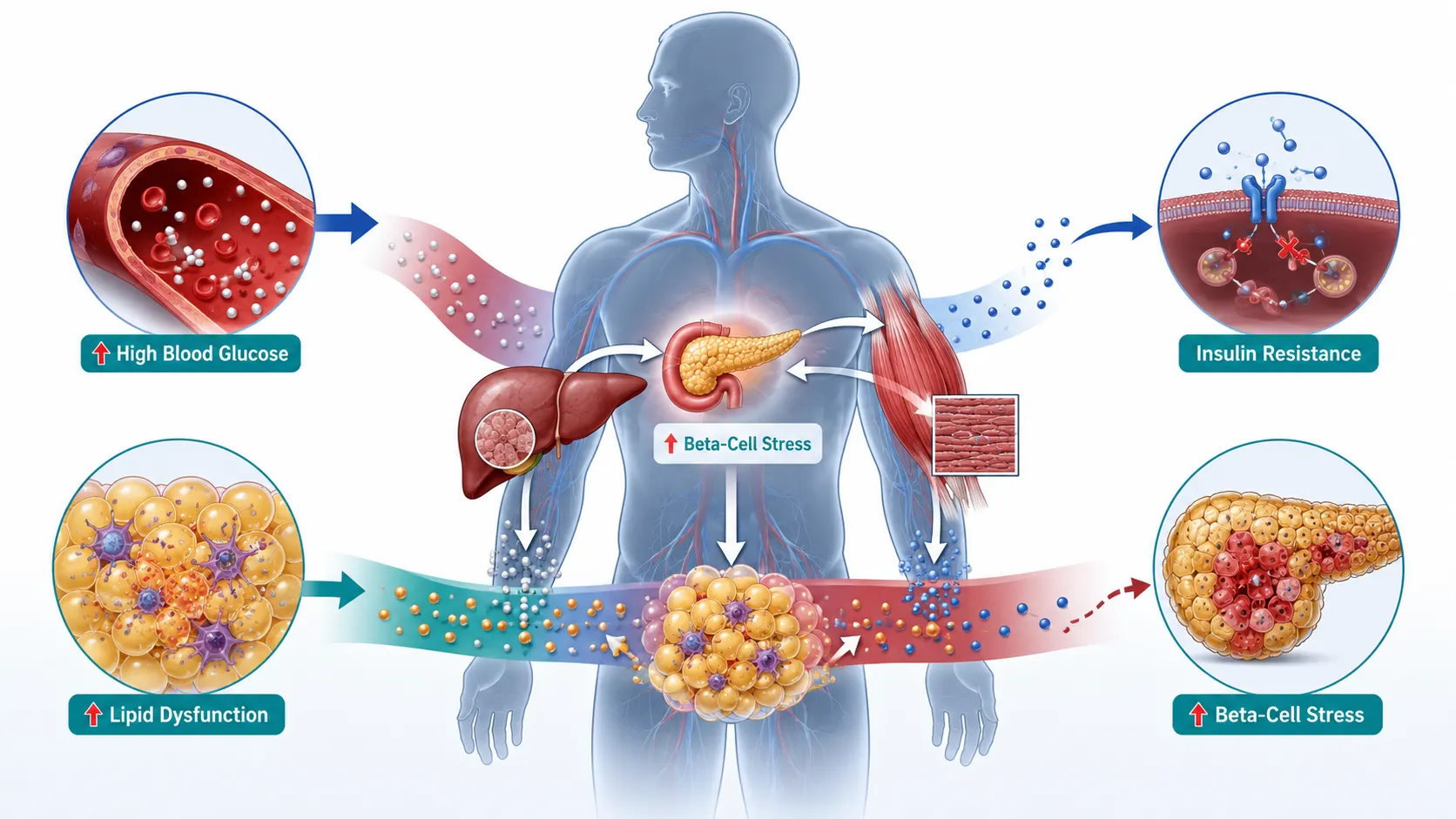
Key Definitions and Concepts
| Term | Definition | Clinical and Biochemical Significance |
|---|---|---|
| Glucose homeostasis | The regulation of blood glucose within a physiologically appropriate range through coordinated hormonal and metabolic processes. | Disruption leads to impaired fasting glucose, impaired glucose tolerance and eventually persistent hyperglycaemia. |
| Insulin resistance | A reduced biological response of target tissues to the actions of insulin. | Often develops in skeletal muscle, liver and adipose tissue before overt type 2 diabetes. |
| Beta-cell dysfunction | Progressive impairment in the ability of pancreatic beta cells to produce and appropriately secrete insulin. | A major factor in the transition from compensated insulin resistance to clinical diabetes. |
| Hepatic glucose production | The production and release of glucose by the liver through glycogenolysis and gluconeogenesis. | Inappropriate elevation contributes significantly to fasting hyperglycaemia. |
| Lipolysis | The breakdown of stored triglycerides into glycerol and free fatty acids. | Excessive release of free fatty acids can worsen insulin resistance and metabolic dysfunction. |
| Lipogenesis | The synthesis and storage of fatty acids and triglycerides. | Dysregulation contributes to ectopic fat accumulation and metabolic disease. |
| Dyslipidaemia | An abnormal pattern of circulating lipids and lipoproteins. | Frequently accompanies insulin resistance and increases cardiovascular risk. |
| Glucotoxicity | Cellular dysfunction resulting from prolonged exposure to elevated glucose concentrations. | Can contribute to further insulin resistance and beta-cell impairment. |
| Lipotoxicity | Cellular damage or dysfunction caused by excessive exposure to fatty acids or lipid intermediates. | May impair insulin signalling and pancreatic beta-cell function. |
| Ectopic fat | Excess lipid accumulation in tissues not primarily designed for long-term fat storage. | Fat accumulation in the liver, skeletal muscle and pancreas may contribute to insulin resistance and organ dysfunction. |
Understanding Normal Glucose and Lipid Metabolism
Before examining metabolic disease, it is necessary to understand how glucose and lipid metabolism normally function. In healthy individuals, multiple organs and hormones work together to maintain energy balance.
Following carbohydrate consumption, glucose enters the bloodstream and stimulates the release of insulin from pancreatic beta cells. Insulin promotes glucose uptake and utilisation in insulin-sensitive tissues, particularly skeletal muscle and adipose tissue. It also influences the liver by suppressing excessive glucose production.
During periods of fasting, insulin concentrations decrease and counter-regulatory processes help maintain an adequate supply of glucose. The liver releases glucose through glycogenolysis and produces glucose through gluconeogenesis.
Normal metabolic regulation depends on coordinated actions involving:
The pancreas, which produces insulin and other regulatory hormones.
The liver, which stores, produces and releases glucose.
Skeletal muscle, which represents a major site of glucose disposal.
Adipose tissue, which stores energy and releases fatty acids when required.
The gastrointestinal system, which influences nutrient absorption and hormonal signalling.
The central nervous system, which contributes to appetite and energy regulation.
Hormonal pathways that coordinate nutrient availability and energy utilisation.
Lipid metabolism is equally important. Dietary and internally synthesised lipids are transported through the circulation in complex lipoprotein particles. Adipose tissue stores triglycerides during periods of energy availability and releases fatty acids during periods of increased energy demand.
In a metabolically healthy state, these processes are flexible and appropriately regulated. Type 2 diabetes develops when this flexibility progressively deteriorates.
The Progressive Nature of Type 2 Diabetes
Type 2 diabetes usually develops through a series of overlapping metabolic stages rather than appearing suddenly. The precise sequence differs between individuals, but a common pattern involves the gradual development of insulin resistance followed by progressive failure of compensatory insulin secretion.
Early Metabolic Susceptibility
Genetic predisposition, body composition, dietary patterns, physical inactivity, ageing and other environmental factors can contribute to metabolic vulnerability. These factors do not affect every individual in the same way.
Important contributors may include:
Increased accumulation of visceral adipose tissue.
Reduced physical activity and skeletal muscle glucose utilisation.
Excess energy intake relative to long-term requirements.
Genetic susceptibility affecting insulin secretion or insulin action.
Chronic low-grade inflammation.
Sleep disruption and altered circadian patterns.
Age-related changes in body composition.
Ectopic accumulation of lipid in metabolically important organs.
These factors interact rather than acting independently. For example, increased adiposity may influence both lipid metabolism and inflammatory signalling, which can subsequently affect insulin sensitivity.
The Compensated Insulin Resistance Stage
Insulin resistance may initially develop while blood glucose remains within a relatively normal range. This is possible because pancreatic beta cells increase insulin secretion to compensate for reduced insulin effectiveness.
At this stage:
Target tissues require more insulin to produce a similar metabolic response.
Circulating insulin concentrations may increase.
Glucose regulation may remain apparently normal.
Lipid abnormalities may already be developing.
Metabolic stress may be increasing despite the absence of overt diabetes.
This stage demonstrates an important clinical principle: normal glucose values do not necessarily indicate complete metabolic health.
The Biochemical Basis of Insulin Resistance
Insulin resistance is central to the progression of type 2 diabetes. It occurs when cells respond less effectively to insulin, requiring higher concentrations of the hormone to produce an appropriate metabolic response.
Insulin Signalling Under Normal Conditions
Insulin binds to receptors on the surface of target cells and initiates a sequence of intracellular signalling events. These pathways influence glucose transport, glycogen synthesis, lipid metabolism and protein metabolism.
In skeletal muscle, an important effect of insulin is the stimulation of glucose transport into cells. The absorbed glucose can then be used for energy production or stored as glycogen.
In the liver, insulin normally suppresses excessive glucose production and supports energy storage after nutrient intake.
In adipose tissue, insulin influences glucose uptake and suppresses excessive breakdown of stored triglycerides.
Disruption of Insulin Signalling
When insulin resistance develops, these actions become less effective. The underlying mechanisms are complex and may involve several interacting biochemical disturbances.
Potential contributors include:
Excess intracellular lipid intermediates.
Altered phosphorylation of insulin signalling proteins.
Inflammatory signalling pathways.
Mitochondrial stress and altered energy metabolism.
Oxidative stress.
Endoplasmic reticulum stress.
Changes in adipokine production.
Chronic exposure to excess nutrients.
The critical point is that insulin resistance is not simply a reduction in insulin concentration. It represents altered cellular responsiveness to insulin.
Skeletal Muscle and Impaired Glucose Disposal
Skeletal muscle is one of the most important tissues involved in post-meal glucose disposal. When muscle becomes insulin resistant, glucose uptake following meals may be reduced.
This creates several consequences:
More glucose remains in the circulation.
The pancreas may increase insulin secretion.
Post-meal glucose concentrations may gradually rise.
Long-term demand on beta cells increases.
Metabolic flexibility may decline.
Example
Consider an individual with developing insulin resistance. After consuming a carbohydrate-containing meal, the pancreas releases insulin. However, skeletal muscle does not respond as efficiently as expected.
The sequence may be:
Blood glucose rises following food intake.
Insulin secretion increases.
Muscle glucose uptake is less efficient.
The pancreas produces additional insulin to compensate.
Blood glucose eventually returns towards baseline, but greater insulin secretion is required.
Repeated metabolic stress may contribute to progressive beta-cell dysfunction.
This example illustrates why insulin resistance can remain clinically silent for a considerable period.
Hepatic Insulin Resistance and Excess Glucose Production
The liver plays a major role in maintaining blood glucose concentrations during fasting. Under normal conditions, insulin helps regulate hepatic glucose output.
In hepatic insulin resistance, suppression of glucose production may become impaired. As a result, the liver may continue producing or releasing glucose even when circulating glucose is already elevated.
Major Hepatic Processes
The liver contributes to blood glucose through:
Glycogenolysis, which involves the breakdown of stored glycogen.
Gluconeogenesis, which involves the production of new glucose from non-carbohydrate precursors.
Inappropriately increased activity of these processes can contribute to fasting hyperglycaemia.
Progressive Effects
As hepatic insulin resistance develops:
Fasting glucose may begin to rise.
Hepatic lipid synthesis may become dysregulated.
Triglyceride accumulation may increase.
Very-low-density lipoprotein production may be altered.
Ectopic liver fat may contribute to further metabolic dysfunction.
This demonstrates the close biochemical relationship between glucose and lipid metabolism.
Adipose Tissue Dysfunction
Adipose tissue is not merely an inactive storage site for body fat. It is metabolically active and produces signalling molecules that influence energy regulation, inflammation and insulin sensitivity.
When adipose tissue becomes dysfunctional, its ability to safely store excess energy may be compromised.
Features of Adipose Tissue Dysfunction
Metabolic dysfunction may involve:
Enlargement of existing adipocytes.
Altered capacity for healthy lipid storage.
Increased release of free fatty acids.
Changes in adipokine production.
Increased inflammatory activity.
Recruitment of immune cells into adipose tissue.
Reduced metabolic flexibility.
These changes can affect other organs, particularly the liver and skeletal muscle.
Free Fatty Acids and the Link Between Lipid and Glucose Dysfunction
One of the most important connections between lipid metabolism and type 2 diabetes involves circulating free fatty acids.
Under normal conditions, fatty acids provide an important energy source. However, persistent elevations in circulating fatty acids may contribute to metabolic dysfunction.
Excessive Lipid Availability Can Lead To:
Increased lipid accumulation in skeletal muscle.
Increased hepatic triglyceride accumulation.
Altered insulin signalling.
Increased production of glucose by the liver.
Metabolic stress within pancreatic beta cells.
Increased oxidative stress in susceptible tissues.
This process helps explain why disturbances in lipid metabolism frequently precede or accompany abnormalities in glucose regulation.
Ectopic Fat Accumulation
Ectopic fat refers to lipid accumulation in organs and tissues where excessive fat storage may interfere with normal function.
Important sites include:
The liver.
Skeletal muscle.
The pancreas.
Other metabolically active tissues.
Why Ectopic Fat Is Important
The location of lipid accumulation can be as clinically significant as the total amount of body fat. Some individuals may have relatively high levels of body fat while maintaining comparatively healthy metabolic function, whereas others may develop substantial metabolic dysfunction associated with ectopic lipid accumulation.
Ectopic fat may contribute to:
Hepatic insulin resistance.
Impaired skeletal muscle glucose uptake.
Altered beta-cell function.
Increased lipid intermediate formation.
Inflammatory and oxidative stress.
Dysregulated Lipid Metabolism in Type 2 Diabetes
Type 2 diabetes is frequently associated with a characteristic pattern of lipid abnormalities. These disturbances are not merely secondary findings; they are closely connected to underlying insulin resistance.
Common Metabolic Changes May Include:
Increased triglyceride concentrations.
Altered production and clearance of lipoprotein particles.
Reduced concentrations of protective high-density lipoprotein cholesterol in some individuals.
Changes in the composition and behaviour of low-density lipoprotein particles.
Increased hepatic triglyceride synthesis.
Greater mobilisation of fatty acids from dysfunctional adipose tissue.
The exact pattern varies between individuals and populations. Clinical interpretation should therefore consider the whole metabolic profile rather than relying on a single lipid value.
The Role of De Novo Lipogenesis
De novo lipogenesis refers to the synthesis of fatty acids from non-lipid substrates, particularly when excess energy and carbohydrate availability contribute to lipid production.
In metabolic dysfunction, increased hepatic lipid synthesis may contribute to triglyceride accumulation.
The process can be influenced by:
Excess energy availability.
Hormonal signalling.
Carbohydrate metabolism.
Insulin-related regulatory pathways.
Genetic and environmental factors.
When lipid production and storage exceed the body’s capacity for healthy metabolic handling, fat may accumulate in the liver and other tissues.
Pancreatic Beta-Cell Compensation
The pancreas initially attempts to overcome insulin resistance through increased insulin production.
This compensatory process is critical during the early stages of metabolic dysfunction.
Beta-Cell Compensation May Involve:
Increased insulin synthesis.
Increased insulin secretion following meals.
Adaptation to rising glucose concentrations.
Increased workload on pancreatic cells.
For a period, this response may successfully maintain relatively normal glucose concentrations.
However, compensation has biological limits.
Progressive Beta-Cell Dysfunction
The transition from insulin resistance to clinical type 2 diabetes is strongly associated with progressive impairment of beta-cell function.
As beta cells become unable to produce sufficient insulin for the degree of insulin resistance, blood glucose concentrations rise.
Several mechanisms may contribute to beta-cell dysfunction.
Glucotoxicity
Persistent exposure to elevated glucose may impair cellular function. This can create a harmful cycle:
Insulin secretion becomes inadequate.
Blood glucose rises.
Elevated glucose places additional stress on beta cells.
Beta-cell function may deteriorate further.
Hyperglycaemia becomes increasingly difficult to control.
Lipotoxicity
Excessive exposure to fatty acids and harmful lipid intermediates may also contribute to cellular dysfunction.
Potential consequences include:
Altered insulin secretion.
Cellular stress.
Mitochondrial dysfunction.
Impaired energy regulation.
Increased susceptibility to damage.
Glucolipotoxicity
In reality, elevated glucose and abnormal lipid exposure often occur together. Their combined effects may be more relevant than considering either disturbance independently.
Progressive Dysglycaemia
The biochemical progression towards type 2 diabetes often involves gradual deterioration in glucose regulation.
A Simplified Progression
Normal glucose regulation.
Insulin resistance with compensatory hyperinsulinaemia.
Impaired post-meal glucose regulation.
Impaired fasting glucose in susceptible individuals.
Increasing beta-cell dysfunction.
Persistent hyperglycaemia consistent with clinical diabetes.
Not every individual follows precisely the same sequence. The rate of progression and dominant metabolic abnormalities can vary considerably.
The Importance of Postprandial Metabolism
Postprandial metabolism refers to the body’s metabolic response after eating. Early abnormalities in glucose regulation may become apparent after meals before fasting glucose becomes persistently elevated.
This is because a meal creates a significant metabolic challenge.
The body must:
Absorb nutrients.
Release insulin appropriately.
Suppress excessive hepatic glucose output.
Increase glucose uptake in peripheral tissues.
Regulate lipid transport and storage.
Failure in any of these processes can contribute to abnormal post-meal glucose and lipid responses.
Inflammation and Metabolic Dysfunction
Chronic low-grade inflammation is frequently associated with insulin resistance and obesity-related metabolic dysfunction.
Inflammatory signalling can influence:
Insulin receptor signalling.
Adipose tissue function.
Lipid mobilisation.
Hepatic metabolism.
Vascular health.
It is important to recognise that inflammation may act as both a contributor to and consequence of metabolic dysfunction.
Potential Sources of Metabolic Inflammation Include:
Dysfunctional adipose tissue.
Altered immune activity.
Cellular stress.
Ectopic lipid accumulation.
Associated chronic conditions.
The relationship is complex and should not be simplified into a single cause-and-effect pathway.
Oxidative Stress and Cellular Dysfunction
Oxidative stress occurs when the balance between oxidant production and antioxidant defence becomes disrupted.
Metabolic dysfunction may increase oxidative stress through:
Excess nutrient availability.
Mitochondrial dysfunction.
Chronic hyperglycaemia.
Abnormal fatty acid metabolism.
Inflammatory activation.
Persistent oxidative stress can influence cellular proteins, lipids and other molecular structures.
In type 2 diabetes, oxidative stress is particularly important because it may contribute to both metabolic dysfunction and long-term complications.
Mitochondrial Function and Energy Metabolism
Mitochondria are central to cellular energy production. They participate in the metabolism of glucose, fatty acids and other energy substrates.
Altered mitochondrial function may influence metabolic flexibility, particularly in tissues such as skeletal muscle.
Potential consequences include:
Reduced capacity to adapt to changing energy demands.
Altered fatty acid oxidation.
Accumulation of metabolic intermediates.
Increased oxidative stress.
Impaired cellular signalling.
However, mitochondrial dysfunction should be viewed as part of a complex metabolic network rather than the sole explanation for type 2 diabetes.
The Interrelationship Between Obesity and Type 2 Diabetes
Obesity and type 2 diabetes are strongly associated, but the relationship is biologically complex. Not all individuals living with obesity develop type 2 diabetes, and type 2 diabetes can occur in individuals who do not meet conventional definitions of obesity.
Risk is influenced by factors including:
Distribution of adipose tissue.
Visceral fat accumulation.
Ectopic fat deposition.
Genetic susceptibility.
Beta-cell reserve.
Physical activity.
Dietary patterns.
Age and other physiological factors.
This highlights the importance of avoiding overly simplistic assumptions based solely on body weight.
Visceral Adiposity and Metabolic Risk
Visceral adipose tissue is located around internal organs within the abdominal cavity. Increased visceral adiposity is commonly associated with greater metabolic risk.
Potential mechanisms include:
Increased fatty acid delivery to the liver.
Altered adipokine signalling.
Greater inflammatory activity.
Increased hepatic triglyceride accumulation.
Worsening insulin resistance.
Clinical assessment should therefore consider metabolic indicators alongside anthropometric measures.
Key Biochemical Markers Relevant to Disease Progression
A variety of laboratory measures can contribute to assessment of glucose and lipid metabolism.
Commonly Relevant Measures Include:
Fasting blood glucose.
Measures reflecting longer-term glycaemic exposure.
Post-meal glucose responses where clinically appropriate.
Fasting insulin in selected research or clinical contexts.
Triglyceride concentrations.
Cholesterol and lipoprotein profiles.
Markers of liver metabolism.
Other indicators relevant to metabolic risk.
No single biomarker provides a complete picture of metabolic health.
Interpreting Biochemical Markers Critically
Laboratory results must be interpreted in context.
Factors that may influence results include:
Acute illness.
Medication use.
Recent dietary intake.
Fasting status where relevant.
Physical activity.
Liver and kidney function.
Biological variation.
Laboratory methodology.
A Critical Assessment Approach Should Include:
Reviewing patterns rather than isolated results.
Comparing results over time where appropriate.
Considering symptoms and clinical history.
Identifying possible confounding factors.
Recognising the limitations of individual biomarkers.
Following appropriate professional and clinical guidelines.
Practical Example: Progressive Metabolic Dysfunction
Consider a hypothetical adult who has gradually become less physically active and developed increased central adiposity.
Stage One
Laboratory findings may initially show:
Normal fasting glucose.
Increased insulin requirements to maintain glucose control.
Mild abnormalities in triglyceride metabolism.
The individual may have no obvious symptoms.
Stage Two
Over time:
Post-meal glucose regulation becomes less efficient.
Hepatic insulin resistance increases.
Liver fat accumulation may become more significant.
Beta cells increase insulin production.
Stage Three
Eventually:
Beta-cell compensation becomes inadequate.
Fasting and/or post-meal glucose concentrations increase.
Persistent hyperglycaemia develops.
Clinical criteria for type 2 diabetes may be met.
This simplified example demonstrates the progressive and interconnected nature of the condition.
Critical Evaluation of the Glucose-Centred Model
Type 2 diabetes is often described primarily as a disorder of high blood glucose. Although hyperglycaemia is clinically important, this description is incomplete.
A glucose-centred model may fail to fully address:
Lipid abnormalities.
Adipose tissue dysfunction.
Hepatic metabolic changes.
Inflammation.
Beta-cell failure.
Ectopic fat.
Individual differences in disease mechanisms.
A more comprehensive understanding recognises type 2 diabetes as a heterogeneous metabolic condition.
Heterogeneity in Type 2 Diabetes
Different individuals may develop type 2 diabetes through somewhat different dominant pathways.
For example:
One person may have pronounced insulin resistance with initially preserved insulin secretion.
Another may develop significant beta-cell dysfunction at a lower level of insulin resistance.
Another may have substantial hepatic metabolic dysfunction.
Another may experience a combination of these processes.
This heterogeneity has important implications for research and personalised clinical care.
Clinical Benefits of Understanding Progressive Biochemical Dysfunction
A strong understanding of the biochemical progression of type 2 diabetes offers several practical benefits.
Key Benefits Include:
Earlier recognition of metabolic risk.
Improved interpretation of glucose and lipid profiles.
Better understanding of disease progression.
More informed nutritional assessment.
Greater ability to evaluate therapeutic interventions.
Improved communication within multidisciplinary teams.
Support for evidence-informed clinical decision-making.
Recognition that metabolic health involves multiple interacting systems.
Applying Biochemical Knowledge in Clinical Practice
Healthcare and nutrition professionals may encounter individuals with complex metabolic profiles. Effective assessment requires structured reasoning.
A Systematic Approach May Include:
Reviewing the individual’s medical and family history.
Assessing relevant dietary and lifestyle information.
Considering body composition and fat distribution where appropriate.
Reviewing glucose-related laboratory findings.
Examining lipid metabolism indicators.
Considering liver and other organ function.
Identifying potential confounding factors.
Evaluating changes over time.
Recognising when specialist assessment is required.
Supporting evidence-based management within the individual’s professional scope of practice.
Practical Scenario: Interpreting Combined Glucose and Lipid Changes
A patient presents with progressively increasing fasting glucose, elevated triglycerides and evidence of increased central adiposity.
A narrow interpretation might focus exclusively on glucose.
A more comprehensive biochemical assessment would consider:
Possible insulin resistance.
Hepatic lipid metabolism.
Adipose tissue dysfunction.
The potential contribution of increased fatty acid flux.
Lifestyle and dietary factors.
Medication use.
Family history and other risk factors.
The combined pattern may provide more useful information than any single result alone.
The Role of Lifestyle in Metabolic Biochemistry
Dietary patterns and physical activity can influence several pathways involved in glucose and lipid metabolism.
Physical activity can affect:
Skeletal muscle glucose utilisation.
Insulin sensitivity.
Energy expenditure.
Lipid oxidation.
Body composition.
Dietary intake can influence:
Postprandial glucose responses.
Energy balance.
Lipid availability.
Hepatic metabolism.
Overall nutritional status.
However, effective intervention requires individual assessment. A single nutritional strategy is unlikely to be equally appropriate for every person.
Key Principles for Learners
Learners should retain several fundamental principles when examining the onset of type 2 diabetes.
Essential Principles
Type 2 diabetes develops progressively in many individuals.
Insulin resistance may occur before persistent hyperglycaemia becomes clinically evident.
Beta-cell compensation can temporarily maintain glucose homeostasis.
Progressive beta-cell dysfunction contributes to clinical disease onset.
Glucose and lipid metabolism are closely interconnected.
Dysfunctional adipose tissue can influence systemic metabolism.
Excess fatty acid exposure may contribute to insulin resistance and cellular dysfunction.
Ectopic fat accumulation can impair the function of metabolically important organs.
Chronic inflammation and oxidative stress may contribute to disease progression.
Individual metabolic pathways and disease trajectories vary.
Laboratory markers should be interpreted as part of a wider clinical assessment.
Summary
The clinical onset of type 2 diabetes represents the culmination of progressive biochemical disturbances rather than a sudden failure of glucose regulation. Early insulin resistance may initially be compensated for through increased insulin secretion, allowing blood glucose concentrations to remain relatively controlled. Over time, however, persistent metabolic stress can impair the ability of pancreatic beta cells to maintain adequate insulin secretion.
Disturbances in lipid metabolism are closely integrated with these processes. Dysfunctional adipose tissue, increased release of fatty acids, hepatic lipid accumulation and ectopic fat deposition can contribute to worsening insulin resistance and cellular dysfunction. These changes demonstrate that glucose and lipid metabolism cannot be considered independently when examining the development of type 2 diabetes.
The liver, skeletal muscle, adipose tissue and pancreas form an interconnected metabolic network. Progressive dysfunction within one system can influence the function of others, creating cycles of worsening insulin resistance, abnormal lipid handling and impaired glucose control. Inflammatory processes, oxidative stress and altered mitochondrial function may further contribute to this complex metabolic environment.
For clinical and nutritional practice, this knowledge supports a more comprehensive approach to metabolic assessment. Rather than focusing solely on a single glucose measurement, professionals should understand broader biochemical patterns and recognise the importance of individual variation. Critically examining the progressive interaction between glucose and lipid metabolism provides an essential scientific foundation for understanding type 2 diabetes, identifying metabolic risk and supporting evidence-informed therapeutic strategies.
2.Evaluate the Specific Molecular Pathways Through Which Chronic Systemic Inflammation Contributes to the Development of Obesity and Broader Metabolic Syndrome
Chronic systemic inflammation is increasingly recognised as an important biological process involved in the development and progression of obesity and metabolic syndrome. Unlike the rapid and often intense inflammatory response associated with acute infection or injury, chronic metabolic inflammation is generally low-grade, persistent and closely linked to long-term disturbances in energy balance, adipose tissue function and metabolic regulation.
Obesity is not simply the result of increased body mass or excess energy storage. In many individuals, particularly those with increased visceral and ectopic adipose tissue, obesity is associated with substantial changes in cellular communication, immune activity, lipid metabolism and hormonal regulation. Enlarged and metabolically stressed adipocytes can alter the production of signalling molecules, recruit immune cells and contribute to systemic inflammatory activity. These processes may impair insulin signalling and influence glucose and lipid metabolism.
Metabolic syndrome refers to a clustering of interconnected metabolic abnormalities that may include central adiposity, impaired glucose regulation, insulin resistance, dyslipidaemia and elevated cardiovascular risk factors. Chronic inflammation does not act independently of these conditions. Instead, it interacts with adipose tissue dysfunction, excess fatty acid availability, oxidative stress and altered endocrine signalling to create self-reinforcing cycles of metabolic dysfunction.
This section critically examines the major molecular pathways involved and explains how persistent inflammatory signalling can influence the development of obesity and broader metabolic syndrome. Learners will develop the ability to connect molecular and cellular mechanisms with clinical metabolic patterns while recognising that obesity and metabolic syndrome are biologically heterogeneous conditions.
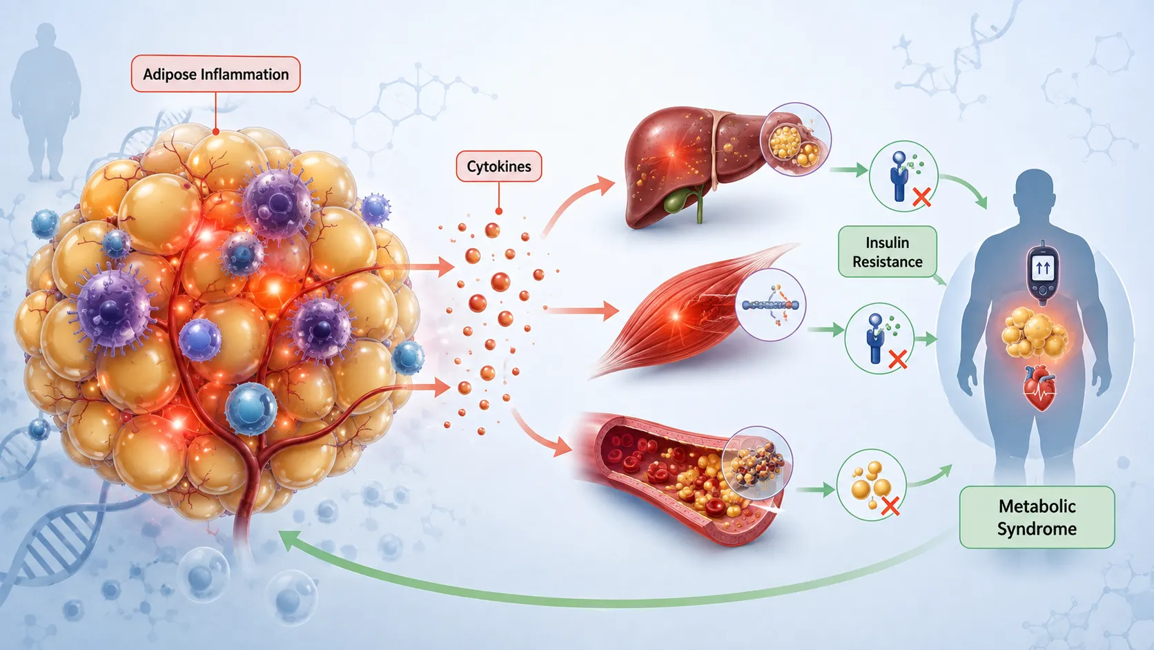
Key Definitions and Concepts
| Term | Definition | Relevance to Obesity and Metabolic Syndrome |
|---|---|---|
| Chronic systemic inflammation | Persistent, generally low-grade activation of inflammatory processes affecting multiple tissues or the wider circulation. | Can interfere with metabolic signalling and contribute to insulin resistance and tissue dysfunction. |
| Metabolic inflammation | Inflammatory activity associated with nutrient excess, adipose tissue dysfunction and metabolic stress. | Links altered energy metabolism with immune signalling. |
| Adipokine | A signalling molecule produced by adipose tissue. | Can influence appetite, inflammation, insulin sensitivity and lipid metabolism. |
| Cytokine | A small signalling protein involved in communication between cells, particularly immune and inflammatory cells. | Certain cytokines can alter insulin signalling and metabolic regulation. |
| Insulin resistance | A reduced response of target tissues to insulin. | A central metabolic abnormality associated with obesity and metabolic syndrome. |
| Oxidative stress | A disturbance between oxidant production and antioxidant defence that may contribute to cellular dysfunction. | Can amplify inflammatory and metabolic signalling disturbances. |
| Visceral adiposity | Accumulation of adipose tissue around internal abdominal organs. | Frequently associated with increased metabolic and inflammatory risk. |
| Lipotoxicity | Cellular dysfunction associated with excessive exposure to fatty acids or harmful lipid intermediates. | Can impair insulin signalling and contribute to organ dysfunction. |
| Ectopic fat | Excess lipid accumulation in tissues not primarily designed for long-term fat storage. | May contribute to inflammation and metabolic dysfunction in organs such as the liver and skeletal muscle. |
| Metabolic syndrome | A cluster of metabolic abnormalities associated with increased cardiometabolic risk. | Chronic inflammation may interact with several components of the syndrome. |
Understanding Chronic Systemic Inflammation
Inflammation is a fundamental protective biological response. Acute inflammation can help the body respond to infection, tissue damage and other immediate threats. Under normal circumstances, inflammatory pathways are activated for an appropriate period and subsequently regulated or resolved.
Chronic systemic inflammation differs because inflammatory activity remains persistent or repeatedly activated over a longer period. In metabolic disease, this process is often described as low-grade because it may not produce the dramatic symptoms associated with severe infection.
Key Characteristics of Chronic Metabolic Inflammation
Chronic metabolic inflammation may involve:
Persistent activation of immune signalling pathways.
Altered production of cytokines and other inflammatory mediators.
Changes in adipose tissue immune cell populations.
Increased oxidative and cellular stress.
Dysregulated lipid metabolism.
Impaired insulin signalling.
Communication between metabolically active organs.
Progressive effects on vascular and organ function.
It is important to recognise that inflammation may be both a consequence and a contributor to metabolic dysfunction.
The Relationship Between Obesity and Inflammation
Adipose tissue is an active endocrine and metabolic organ. It stores energy, releases fatty acids and produces a range of signalling molecules. When adipose tissue expands beyond its capacity for healthy adaptation, several structural and biochemical changes may occur.
Adipocytes may enlarge, local oxygen availability may change, extracellular tissue structures may remodel and immune cells may become increasingly involved in the local tissue environment. These changes can promote inflammatory signalling.
Adipose Tissue Expansion Can Involve
Adipocyte enlargement.
Altered adipocyte development.
Changes in local blood supply.
Tissue hypoxia in susceptible areas.
Increased cellular stress.
Altered adipokine secretion.
Immune cell recruitment.
Changes in extracellular matrix structure.
Not all adipose tissue expansion produces the same metabolic consequences. The location, biological characteristics and functional capacity of adipose tissue are important.
Adipocyte Hypertrophy and Cellular Stress
When adipocytes become enlarged, their metabolic demands and physical environment may change. Large adipocytes can experience increased stress related to nutrient handling, oxygen availability and intracellular lipid storage.
This cellular stress may activate signalling pathways that influence inflammatory mediator production.
Potential Consequences Include
Altered mitochondrial activity.
Endoplasmic reticulum stress.
Increased oxidative stress.
Changes in cytokine production.
Altered adipokine release.
Increased lipolytic activity in some metabolic states.
Impaired insulin responsiveness.
These disturbances may transform adipose tissue from an efficient energy-storage organ into a contributor to systemic metabolic dysfunction.
Immune Cell Infiltration of Adipose Tissue
One of the important features of metabolically dysfunctional adipose tissue is a change in the immune cell environment.
As adipose tissue becomes stressed, signalling molecules may attract immune cells. Macrophages are particularly important in many models of metabolic inflammation, although several immune cell populations can participate.
Immune Changes May Include
Increased macrophage accumulation.
Altered macrophage functional states.
Changes in T-cell activity.
Activation of other innate immune pathways.
Increased production of inflammatory mediators.
Altered communication between immune cells and adipocytes.
The result can be a local inflammatory environment that affects both adipose tissue and systemic metabolism.
Cytokines and Metabolic Regulation
Cytokines are signalling molecules that regulate communication between cells. In chronic inflammation, altered cytokine activity can influence multiple tissues.
Certain pro-inflammatory cytokines are associated with pathways that may impair insulin action and alter metabolic regulation. Their effects depend on concentration, duration, tissue context and interactions with other biological processes.
Important Effects of Inflammatory Cytokine Signalling May Include
Alteration of insulin receptor signalling.
Changes in glucose uptake.
Modification of lipid metabolism.
Increased hepatic inflammatory activity.
Effects on endothelial and vascular function.
Stimulation of additional inflammatory pathways.
A critical point is that cytokines have complex biological roles. They should not be viewed simply as harmful molecules, because many also participate in normal immune regulation and tissue repair.
Nuclear Factor Kappa B and Inflammatory Gene Expression
One important molecular pathway involved in inflammatory signalling is the nuclear factor kappa B pathway, commonly abbreviated as NF-κB.
NF-κB acts as a transcriptional regulator that can influence the expression of multiple genes involved in immune and inflammatory responses.
When inflammatory or cellular stress signals activate this pathway, NF-κB can contribute to the production of additional inflammatory mediators.
General Sequence of Activation
A simplified pathway may involve:
Detection of a metabolic or inflammatory stress signal.
Activation of intracellular signalling processes.
Release or activation of NF-κB-related transcriptional activity.
Movement of activated signalling components towards the cell nucleus.
Alteration of inflammatory gene expression.
Production of signalling molecules that may influence neighbouring or distant tissues.
Persistent activation may contribute to the maintenance of chronic inflammatory activity.
Inflammation and Insulin Signalling
The connection between inflammation and insulin resistance is one of the most important mechanisms linking obesity to metabolic syndrome.
Under normal conditions, insulin initiates intracellular signalling pathways that promote appropriate glucose uptake, storage and metabolic regulation.
Inflammatory pathways can interfere with elements of this signalling network.
Potential Mechanisms Include
Altered phosphorylation of insulin-signalling proteins.
Activation of stress-related kinases.
Increased production of inflammatory mediators.
Accumulation of harmful lipid intermediates.
Oxidative stress.
Endoplasmic reticulum stress.
These mechanisms can reduce the efficiency with which cells respond to insulin.
Stress Kinases and Metabolic Dysfunction
Several intracellular stress-response pathways can be activated by inflammatory mediators, oxidative stress and excess nutrient availability.
Stress kinases may influence insulin signalling by modifying proteins involved in metabolic communication.
Their Activation May Be Associated With
Chronic nutrient excess.
Increased free fatty acid exposure.
Inflammatory cytokines.
Reactive oxygen species.
Cellular stress.
Ectopic lipid accumulation.
Persistent activation may contribute to reduced insulin sensitivity in skeletal muscle, liver and adipose tissue.
The Role of Adipokines
Adipose tissue produces several biologically active molecules known as adipokines. These molecules contribute to communication between adipose tissue and other organs.
In metabolically healthy adipose tissue, adipokine production can support normal metabolic regulation. During adipose tissue dysfunction, the balance of these signals may change.
Adipokine Dysregulation May Affect
Insulin sensitivity.
Appetite regulation.
Energy expenditure.
Inflammatory activity.
Lipid metabolism.
Vascular function.
The overall metabolic effect depends on the combined pattern of adipose tissue signalling rather than a single molecule.
Visceral Adiposity and Inflammatory Risk
Visceral adipose tissue is particularly important when examining metabolic syndrome. It is located within the abdominal cavity around internal organs and has distinct metabolic characteristics.
Increased visceral adiposity is frequently associated with:
Greater metabolic inflammation.
Increased fatty acid delivery to the liver.
Insulin resistance.
Dyslipidaemia.
Altered hepatic metabolism.
Increased cardiometabolic risk.
Why Location Matters
Two individuals with a similar total body weight may have very different metabolic risk profiles because of differences in:
Visceral fat accumulation.
Subcutaneous adipose tissue capacity.
Ectopic fat distribution.
Genetic susceptibility.
Muscle mass.
Physical activity.
Underlying metabolic health.
Therefore, body weight alone does not provide a complete assessment of metabolic risk.
Free Fatty Acids and Inflammatory Activation
Adipose tissue normally releases fatty acids to provide energy during periods of increased metabolic demand. However, excessive or poorly regulated fatty acid release may contribute to systemic metabolic stress.
Elevated free fatty acid availability can influence both lipid and inflammatory pathways.
Potential Effects Include
Increased hepatic triglyceride synthesis.
Accumulation of lipid intermediates.
Altered insulin signalling.
Activation of cellular stress pathways.
Increased oxidative stress.
Changes in inflammatory mediator production.
This creates a close relationship between lipotoxicity and inflammation.
Molecular Recognition Pathways and Metabolic Stress
Cells possess systems that recognise signals associated with infection, tissue injury and cellular stress. Some of these molecular recognition pathways can also respond to metabolic disturbances.
Excessive nutrient availability and altered lipid environments may contribute to activation of pathways that promote inflammatory responses.
The exact mechanisms remain an active area of scientific research and may vary between tissues and experimental models.
A Simplified Metabolic Inflammation Cycle
Excess nutrient availability increases metabolic stress.
Adipose tissue expands and becomes dysfunctional.
Cellular stress and altered lipid release develop.
Immune and inflammatory pathways become activated.
Cytokine and stress signalling interfere with metabolic regulation.
Insulin resistance worsens.
Further nutrient and lipid abnormalities reinforce the cycle.
This model illustrates why chronic inflammation can contribute to progressive metabolic disease.
Oxidative Stress as a Molecular Amplifier
Oxidative stress can interact closely with inflammation. Reactive oxygen species are produced during normal cellular metabolism and can serve useful biological signalling roles.
Problems arise when oxidant production exceeds the capacity of protective systems.
Metabolic Conditions That May Increase Oxidative Stress Include
Persistent hyperglycaemia.
Excessive fatty acid availability.
Mitochondrial dysfunction.
Chronic inflammation.
Ectopic lipid accumulation.
Oxidative stress can activate additional inflammatory pathways and contribute to cellular damage.
The Inflammation–Oxidative Stress Cycle
Chronic inflammation may increase oxidative stress, while oxidative stress can activate further inflammatory signalling. This creates a reinforcing biological cycle.
The consequences may include:
Reduced insulin sensitivity.
Altered endothelial function.
Cellular damage.
Mitochondrial stress.
Progressive metabolic dysfunction.
Mitochondrial Dysfunction and Inflammation
Mitochondria are essential for energy production and metabolic flexibility. In obesity and metabolic syndrome, altered mitochondrial function may contribute to changes in substrate utilisation.
Mitochondrial dysfunction may lead to:
Reduced efficiency of energy metabolism.
Altered fatty acid oxidation.
Increased reactive oxygen species.
Accumulation of metabolic intermediates.
Activation of stress signalling pathways.
Mitochondria can therefore connect nutrient metabolism with inflammatory activity.
However, it is important not to assume that mitochondrial dysfunction is always the initial cause. In some situations, it may develop as a consequence of other metabolic disturbances.
Endoplasmic Reticulum Stress
The endoplasmic reticulum is involved in protein processing, lipid metabolism and cellular signalling. Excess nutrient exposure can place pressure on this system.
When cells experience persistent metabolic overload, endoplasmic reticulum stress responses may become activated.
Potential Consequences Include
Altered protein processing.
Activation of stress-response pathways.
Changes in inflammatory signalling.
Impaired insulin action.
Increased cellular dysfunction.
This pathway provides another mechanism through which nutrient excess may become linked to inflammation.
The NLRP3 Inflammasome and Metabolic Inflammation
The inflammasome is a multi-protein signalling system involved in aspects of innate immune regulation. Certain inflammasome pathways have been investigated in relation to metabolic inflammation.
The NLRP3 inflammasome is frequently discussed in research concerning obesity and metabolic dysfunction.
Its Potential Activation May Be Influenced By
Cellular stress.
Oxidative stress.
Altered lipid metabolism.
Metabolic danger signals.
Mitochondrial dysfunction.
Activation can contribute to the maturation and release of specific inflammatory mediators.
Critical Evaluation
Although experimental and clinical research has highlighted the potential importance of inflammasome signalling, the precise contribution of these pathways can vary. Learners should avoid assuming that inhibition of a single pathway would universally resolve metabolic disease.
Chronic Inflammation and Hepatic Metabolism
The liver is a central organ in glucose and lipid regulation. Chronic systemic inflammation can influence hepatic metabolism in several ways.
Potential Hepatic Effects Include
Increased insulin resistance.
Altered glucose production.
Increased triglyceride synthesis.
Changes in lipid transport.
Increased inflammatory signalling.
Greater accumulation of ectopic fat.
When hepatic insulin resistance develops, the liver may continue producing glucose even when circulating glucose is elevated.
At the same time, dysregulated lipid metabolism can contribute to increased triglyceride production and altered lipoprotein patterns.
This combination links chronic inflammation with both impaired glucose regulation and dyslipidaemia.
Inflammation, Dyslipidaemia and Metabolic Syndrome
Metabolic syndrome commonly includes disturbances in lipid metabolism. Chronic inflammation can interact with these abnormalities through effects on the liver, adipose tissue and vascular system.
Common Patterns May Include
Increased triglyceride concentrations.
Altered lipoprotein metabolism.
Changes in protective lipoprotein profiles.
Increased fatty acid flux.
Hepatic triglyceride accumulation.
These abnormalities can contribute to a broader cardiometabolic risk profile.
Skeletal Muscle Inflammation and Insulin Resistance
Skeletal muscle is a major site of glucose utilisation. Inflammatory signalling and lipid accumulation within muscle can interfere with insulin responsiveness.
Potential mechanisms include:
Accumulation of intracellular lipid intermediates.
Activation of stress kinases.
Oxidative stress.
Altered mitochondrial function.
Changes in inflammatory signalling.
Reduced skeletal muscle insulin sensitivity can substantially affect whole-body glucose regulation because muscle normally accounts for a significant proportion of insulin-stimulated glucose disposal.
Inflammation and the Development of Hyperglycaemia
As inflammation contributes to insulin resistance in the liver, muscle and adipose tissue, the pancreas may initially compensate by increasing insulin secretion.
Over time, this compensation may become inadequate.
A progressive sequence may involve:
Chronic metabolic stress develops.
Inflammatory pathways become persistently activated.
Insulin sensitivity declines.
The pancreas increases insulin secretion.
Beta cells experience increasing metabolic demand.
Glucose regulation deteriorates.
Persistent hyperglycaemia may develop.
Inflammation is therefore part of a wider network that can contribute to the progression from obesity-associated insulin resistance to type 2 diabetes.
Inflammation and Pancreatic Beta-Cell Stress
Persistent exposure to elevated glucose, fatty acids and inflammatory mediators may contribute to beta-cell stress.
Potential consequences include:
Altered insulin synthesis.
Impaired insulin secretion.
Increased oxidative stress.
Mitochondrial dysfunction.
Cellular stress responses.
As beta-cell function declines, the body’s ability to compensate for insulin resistance decreases.
This illustrates how systemic metabolic inflammation can affect multiple organs simultaneously.
The Role of the Gut and Metabolic Inflammation
The gastrointestinal system has an important relationship with metabolism and immune regulation. Dietary patterns, intestinal function and microbial metabolites may influence metabolic signalling.
Research in this area is developing rapidly, and learners should interpret emerging findings carefully.
Potential areas of interaction include:
Gut barrier function.
Immune signalling.
Microbial metabolites.
Dietary fibre metabolism.
Systemic inflammatory responses.
Critical Consideration
The gut microbiome is a highly complex ecosystem. Associations between specific microbial patterns and obesity do not automatically demonstrate direct causation. Research findings must be evaluated according to study design, population characteristics and methodological quality.
Molecular Pathways as Interconnected Networks
A major challenge when studying chronic inflammation is the tendency to examine individual pathways separately. In reality, metabolic regulation involves interconnected molecular networks.
For example:
Excess adipose tissue can alter fatty acid release.
Fatty acids can contribute to cellular stress.
Cellular stress can activate inflammatory pathways.
Inflammation can impair insulin signalling.
Insulin resistance can worsen lipid abnormalities.
Lipid abnormalities can increase ectopic fat.
Ectopic fat can produce further metabolic stress.
This interconnected model is more accurate than a simple one-direction pathway.
Practical Example: The Development of Metabolic Dysfunction
Consider an adult who experiences gradual increases in visceral adiposity over several years.
Initial Stage
The individual may develop:
Increased adipocyte size.
Greater lipid storage demands.
Mild changes in adipokine signalling.
Early insulin resistance.
Blood glucose may remain within a clinically acceptable range because the pancreas compensates with increased insulin secretion.
Progressive Stage
Over time:
Adipose tissue dysfunction becomes more pronounced.
Free fatty acid release increases.
Inflammatory signalling becomes more persistent.
Hepatic lipid accumulation develops.
Skeletal muscle insulin sensitivity declines.
Laboratory findings may begin to demonstrate abnormalities in glucose and lipid metabolism.
Advanced Metabolic Stage
If dysfunction continues:
Beta-cell compensation may become inadequate.
Persistent hyperglycaemia may develop.
Dyslipidaemia may worsen.
Cardiometabolic risk factors may cluster.
This example demonstrates the progressive relationship between inflammation, obesity and metabolic syndrome.
Clinical and Practical Importance of Molecular Understanding
Understanding molecular pathways helps professionals interpret why metabolic disorders are complex and why interventions may have different effects between individuals.
Key Benefits Include
Improved understanding of insulin resistance.
Better interpretation of glucose and lipid abnormalities.
Recognition of the role of adipose tissue distribution.
Greater appreciation of metabolic heterogeneity.
Support for evidence-based therapeutic strategies.
Improved communication within multidisciplinary teams.
Stronger ability to evaluate emerging scientific research.
Assessing Inflammation in Clinical Context
Clinical assessment should not assume that a single inflammatory marker provides a complete picture of metabolic inflammation.
Inflammatory biomarkers may be influenced by:
Acute infection.
Chronic disease.
Physical injury.
Medication use.
Smoking.
Other lifestyle factors.
Laboratory variation.
A Systematic Assessment Approach Should Include
Reviewing the clinical history.
Considering symptoms and comorbidities.
Evaluating body composition and fat distribution where appropriate.
Reviewing glucose and lipid markers.
Considering inflammatory findings in context.
Identifying possible acute causes of inflammation.
Monitoring trends when clinically appropriate.
Recognising the limitations of individual biomarkers.
Referring or escalating complex cases according to professional responsibilities.
Critical Evaluation of Cause and Effect
One of the most important academic skills in this topic is distinguishing association from causation.
Obesity is associated with chronic inflammation, and inflammation is associated with metabolic dysfunction. However, the direction of these relationships can be complex.
Learners Should Consider
Does obesity initiate inflammatory activity?
Can inflammation contribute to weight gain and altered energy regulation?
Are both processes influenced by shared underlying factors?
Do genetic factors influence inflammatory responses?
Does the pathway differ between individuals?
The most scientifically responsible position is to recognise that obesity and chronic inflammation often influence each other through multiple feedback mechanisms.
Limitations in Current Research
Research on metabolic inflammation includes evidence from cell studies, animal models, observational studies and clinical trials. Each type of evidence has strengths and limitations.
Common Limitations Include
Difficulty establishing long-term causation.
Biological differences between experimental models and humans.
Variation in definitions of metabolic health.
Heterogeneity between patient populations.
Challenges measuring local tissue inflammation.
Confounding lifestyle and environmental factors.
Differences in laboratory methods.
These limitations should be considered when interpreting claims about individual molecular pathways.
Workplace and Professional Application
Professionals working in clinical nutrition, healthcare and related fields should understand the scientific basis of metabolic inflammation while remaining within their scope of practice.
Appropriate professional actions include:
Recognising patterns consistent with metabolic risk.
Supporting comprehensive nutritional assessment.
Using evidence-based guidance.
Avoiding oversimplified explanations of obesity.
Communicating scientific concepts clearly.
Working within multidisciplinary teams.
Referring complex cases appropriately.
A molecular understanding should support better clinical reasoning rather than encourage unsupported diagnostic conclusions.
Key Learning Points
Learners should be able to identify the following principles.
Chronic Systemic Inflammation
Is typically persistent and low-grade in metabolic disease.
Can develop through interactions between nutrient excess, cellular stress and immune signalling.
Influences multiple metabolically active organs.
May both contribute to and result from metabolic dysfunction.
Adipose Tissue Dysfunction
Involves more than increased fat storage.
Can include adipocyte enlargement and altered immune activity.
May change adipokine and cytokine production.
Can influence systemic lipid and glucose metabolism.
Molecular Signalling
NF-κB-related pathways can influence inflammatory gene expression.
Stress-response pathways can interfere with insulin signalling.
Oxidative and endoplasmic reticulum stress can amplify dysfunction.
Inflammasome pathways are important areas of ongoing research.
Metabolic Consequences
Insulin resistance can develop in muscle, liver and adipose tissue.
Dysregulated lipid metabolism can worsen inflammatory activity.
Hepatic metabolic dysfunction contributes to glucose and lipid abnormalities.
Chronic metabolic stress may eventually affect pancreatic beta-cell function.
Summary
Chronic systemic inflammation represents an important biological mechanism connecting obesity with broader metabolic dysfunction. Metabolic inflammation develops through complex interactions between adipose tissue expansion, immune activity, altered lipid handling, cellular stress and disrupted hormonal signalling.
As adipose tissue becomes metabolically dysfunctional, adipocytes and immune cells can produce altered patterns of cytokines and adipokines. These signals may activate intracellular pathways that influence inflammatory gene expression and interfere with insulin signalling. NF-κB-related activity, stress kinases, oxidative stress, endoplasmic reticulum stress and inflammasome pathways are among the molecular mechanisms investigated in this process.
The consequences extend beyond adipose tissue. Increased free fatty acid availability and inflammatory signalling can influence skeletal muscle, the liver and pancreatic beta cells. This may reduce insulin sensitivity, alter glucose production, disrupt lipid metabolism and contribute to progressive metabolic syndrome.
Importantly, chronic inflammation should not be viewed as a single independent cause of obesity or metabolic syndrome. The relationship is multidirectional. Obesity can promote inflammatory activity, while inflammation may contribute to further disturbances in energy and metabolic regulation. Genetic, behavioural, environmental and physiological factors also influence individual risk.
A critical understanding of these molecular pathways enables Learners to interpret obesity and metabolic syndrome as complex, interconnected disorders. It supports evidence-based clinical reasoning, improved interpretation of metabolic biomarkers and a more comprehensive understanding of therapeutic strategies aimed at reducing long-term metabolic risk.
3.Analyse Complex Clinical and Laboratory Data to Accurately Trace the Biochemical Progression of Insulin Resistance at Both the Cellular and Tissue Levels
Insulin resistance is a progressive metabolic disturbance in which target cells and tissues demonstrate a reduced biological response to insulin. It is a central feature of several metabolic disorders, including obesity, metabolic syndrome and type 2 diabetes. However, insulin resistance is not a single event that can be identified through one laboratory result. It develops through a complex interaction of molecular, cellular, tissue-specific and whole-body metabolic changes.
The accurate analysis of insulin resistance therefore requires the integration of clinical information with biochemical and laboratory data. A meaningful assessment considers factors such as fasting and postprandial glucose, insulin concentrations, glycated markers, lipid profiles, body fat distribution, liver function and indicators of systemic metabolic stress. These findings must then be interpreted in relation to the underlying mechanisms operating within skeletal muscle, adipose tissue, the liver and pancreatic beta cells.
At the cellular level, insulin resistance involves disruption of the normal insulin-signalling cascade. At the tissue level, the consequences differ according to the metabolic function of each organ. Skeletal muscle may show reduced glucose uptake, the liver may demonstrate impaired suppression of glucose production, and adipose tissue may exhibit altered lipid storage and increased fatty acid release. Over time, compensatory increases in insulin secretion may become insufficient, allowing dysglycaemia and progressive metabolic disease to emerge.
This section explains how complex clinical and laboratory information can be systematically analysed to trace the biochemical progression of insulin resistance from early cellular dysfunction to advanced tissue and whole-body metabolic impairment.
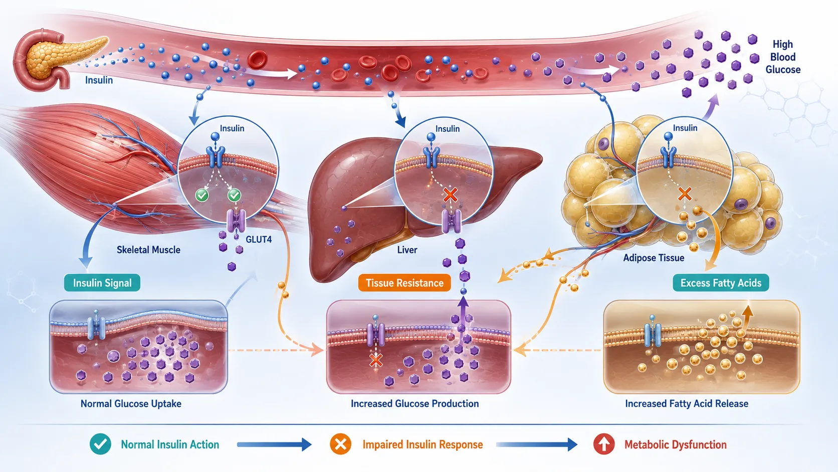
Key Definitions and Concepts
| Term | Definition | Clinical and Biochemical Significance |
|---|---|---|
| Insulin resistance | A reduced biological response of cells or tissues to the actions of insulin. | May precede significant hyperglycaemia and contribute to metabolic syndrome and type 2 diabetes. |
| Hyperinsulinaemia | An elevated circulating insulin concentration, often reflecting increased pancreatic insulin secretion in response to reduced insulin sensitivity. | May occur during compensatory stages of insulin resistance. |
| Insulin sensitivity | The degree to which tissues respond appropriately to insulin. | Reduced sensitivity indicates increasing metabolic dysfunction. |
| Glucose homeostasis | The regulation of blood glucose within an appropriate physiological range. | Becomes progressively impaired as insulin resistance advances. |
| Compensatory insulin secretion | Increased insulin production by pancreatic beta cells to maintain glucose regulation. | Can temporarily maintain normal glucose despite insulin resistance. |
| Hepatic insulin resistance | Reduced responsiveness of the liver to insulin. | May contribute to inappropriate hepatic glucose production and altered lipid metabolism. |
| Peripheral insulin resistance | Reduced insulin responsiveness in tissues such as skeletal muscle and adipose tissue. | Can reduce glucose uptake and alter lipid handling. |
| Lipotoxicity | Cellular dysfunction associated with excessive exposure to fatty acids or harmful lipid intermediates. | Can interfere with insulin signalling and tissue function. |
| Glucotoxicity | Metabolic damage associated with prolonged exposure to elevated glucose concentrations. | May worsen beta-cell function and insulin resistance. |
| Ectopic fat | Excess lipid accumulation in tissues not primarily intended for long-term fat storage. | May contribute to liver and skeletal muscle insulin resistance. |
| HOMA-based assessment | A mathematical estimate using fasting glucose and insulin measurements to approximate insulin resistance or beta-cell function. | Useful in some settings but has important limitations. |
Understanding the Biochemical Progression of Insulin Resistance
Insulin resistance should be understood as a continuum rather than a simple normal-or-abnormal condition. In the earliest stages, cells may become less responsive to insulin while blood glucose remains within an apparently normal range. This occurs because the pancreas can increase insulin secretion to compensate.
As the disturbance progresses, greater amounts of insulin may be required to produce the same metabolic effect. Eventually, pancreatic compensation may become insufficient. At this stage, measurable abnormalities in glucose regulation can emerge.
A Simplified Progression May Include
Early cellular impairment in insulin signalling.
Reduced insulin responsiveness in target tissues.
Increased compensatory insulin secretion.
Development of hyperinsulinaemia.
Altered lipid metabolism.
Increased hepatic glucose production.
Reduced skeletal muscle glucose uptake.
Progressive impairment of glucose tolerance.
Deterioration in beta-cell compensatory capacity.
Persistent hyperglycaemia and possible progression towards type 2 diabetes.
The exact sequence and speed of progression vary substantially between individuals.
Normal Insulin Signalling at the Cellular Level
To understand insulin resistance, it is necessary to understand the normal action of insulin.
Insulin is a peptide hormone produced by pancreatic beta cells. It plays a major role in coordinating nutrient metabolism. Following food intake, increased circulating glucose stimulates insulin secretion, which signals target tissues to regulate glucose uptake, storage and utilisation.
Simplified Cellular Insulin-Signalling Process
A normal sequence broadly involves:
Insulin binds to its receptor on the cell surface.
The insulin receptor undergoes activation.
Intracellular signalling proteins are recruited.
Signalling pathways transmit the metabolic message.
Tissue-specific metabolic responses occur.
Glucose and lipid metabolism are appropriately regulated.
In skeletal muscle and adipose tissue, one important response is the movement of glucose transport mechanisms towards the cell membrane, allowing glucose uptake.
Cellular Disruption in Insulin Resistance
Insulin resistance develops when elements of this signalling network become impaired.
The insulin receptor may still bind insulin, but downstream signalling can become less efficient. As a result, a greater insulin concentration may be required to achieve a comparable metabolic response.
Potential Cellular Mechanisms Include
Altered insulin receptor signalling.
Dysfunction of intracellular signalling proteins.
Activation of stress-related kinases.
Accumulation of lipid intermediates.
Oxidative stress.
Mitochondrial dysfunction.
Endoplasmic reticulum stress.
Chronic inflammatory signalling.
These mechanisms often overlap rather than occurring independently.
The Role of Insulin Receptor Signalling
The insulin receptor initiates a series of intracellular events that communicate the presence of insulin to metabolic pathways within the cell.
When this signalling system is functioning efficiently, insulin can promote appropriate metabolic responses.
During insulin resistance, downstream signalling may become disrupted.
Consequences Can Include
Reduced glucose uptake.
Reduced glycogen synthesis.
Altered lipid metabolism.
Impaired suppression of hepatic glucose production.
Increased reliance on compensatory insulin secretion.
The degree of impairment can vary between tissues.
Phosphorylation and Signal Disruption
Protein phosphorylation is an important mechanism through which cells regulate signalling activity. In metabolic signalling pathways, the location and pattern of phosphorylation can influence whether a protein is activated or inhibited.
Chronic inflammatory and stress pathways can modify key signalling proteins in ways that reduce the effectiveness of insulin signalling.
Factors That May Promote Signalling Disruption Include
Chronic inflammation.
Excess fatty acid exposure.
Cellular oxidative stress.
Persistent nutrient excess.
Ectopic lipid accumulation.
Stress-activated intracellular pathways.
This provides an important molecular link between obesity-associated metabolic stress and insulin resistance.
Lipid Intermediates and Cellular Insulin Resistance
Excess circulating fatty acids can enter tissues such as skeletal muscle and the liver. When the capacity to safely oxidise or store these lipids is exceeded, potentially harmful lipid intermediates may accumulate.
These intermediates can influence intracellular signalling pathways.
Potential Consequences Include
Activation of stress-response proteins.
Impaired insulin signalling.
Mitochondrial stress.
Increased oxidative stress.
Altered cellular energy metabolism.
This process is often described within the broader concept of lipotoxicity.
Oxidative Stress and Insulin Resistance
Reactive oxygen species are produced during normal cellular metabolism. At controlled levels, they participate in physiological signalling. Excessive production, however, can contribute to cellular dysfunction.
Oxidative stress may increase during conditions of:
Chronic hyperglycaemia.
Excessive fatty acid availability.
Mitochondrial dysfunction.
Chronic inflammation.
Persistent metabolic overload.
Effects on Insulin Signalling May Include
Modification of cellular proteins.
Activation of stress kinases.
Altered mitochondrial activity.
Increased inflammatory signalling.
Reduced metabolic flexibility.
Oxidative stress can therefore amplify existing insulin resistance.
Endoplasmic Reticulum Stress
The endoplasmic reticulum plays a central role in protein processing and aspects of lipid metabolism. Persistent nutrient excess can place stress on this cellular system.
When the endoplasmic reticulum becomes overwhelmed, adaptive stress responses may be activated.
Prolonged Stress May Contribute To
Inflammatory signalling.
Altered protein function.
Metabolic dysfunction.
Reduced insulin responsiveness.
Increased cellular injury.
This pathway illustrates the close relationship between cellular nutrient overload and metabolic disease.
Tissue-Specific Progression of Insulin Resistance
Insulin resistance does not affect all tissues in exactly the same way. Different organs have distinct metabolic roles.
The major tissues involved include:
Skeletal muscle.
Liver.
Adipose tissue.
Pancreatic beta cells.
Analysing clinical and laboratory data requires an understanding of these tissue-specific patterns.
Skeletal Muscle Insulin Resistance
Skeletal muscle is an important site of insulin-mediated glucose disposal.
After eating, insulin normally promotes glucose uptake by muscle cells and supports glycogen synthesis.
When skeletal muscle becomes insulin resistant, glucose uptake becomes less efficient.
Biochemical Consequences Include
Reduced insulin-stimulated glucose uptake.
Reduced glycogen synthesis.
Increased circulating glucose after meals.
Increased demand for compensatory insulin secretion.
Altered utilisation of fatty acids and glucose.
Clinical Significance
Skeletal muscle insulin resistance may contribute to abnormal postprandial glucose regulation before fasting glucose becomes clearly abnormal.
Glucose Transport and Cellular Uptake
Insulin helps regulate the movement of glucose transport proteins to the cell membrane in certain insulin-responsive tissues.
When insulin signalling is impaired, this movement may be reduced.
The Result May Be
Glucose remains in the circulation for longer.
Muscle glucose uptake decreases.
The pancreas secretes additional insulin.
Circulating insulin increases.
Insulin resistance may progressively worsen.
This process helps explain why hyperinsulinaemia can develop before persistent hyperglycaemia.
Hepatic Insulin Resistance
The liver plays a major role in maintaining blood glucose during fasting.
Normally, insulin helps suppress excessive hepatic glucose production when circulating glucose and insulin levels rise.
In hepatic insulin resistance, this suppression may become impaired.
Consequences May Include
Continued hepatic glucose production.
Increased fasting glucose.
Increased gluconeogenic activity.
Altered glycogen metabolism.
Increased triglyceride synthesis.
Hepatic insulin resistance therefore contributes to both glucose and lipid abnormalities.
Selective Hepatic Insulin Resistance
One complex feature of hepatic insulin resistance is that not all insulin-related pathways necessarily become resistant to the same degree.
Certain processes may remain responsive while others become impaired.
This can contribute to a combination of:
Inadequate suppression of glucose production.
Continued or altered lipid synthesis.
Increased triglyceride accumulation.
This complexity demonstrates why insulin resistance cannot always be described as a complete loss of insulin action.
Adipose Tissue Insulin Resistance
Insulin normally helps regulate lipid storage and suppress excessive breakdown of stored triglycerides.
When adipose tissue becomes insulin resistant, the ability of insulin to regulate these processes may decline.
Potential Consequences Include
Increased lipolysis.
Increased release of free fatty acids.
Greater fatty acid delivery to the liver.
Ectopic fat accumulation.
Increased metabolic inflammation.
This can create a self-reinforcing cycle between adipose tissue dysfunction and systemic insulin resistance.
Free Fatty Acid Flux
Increased release of free fatty acids from adipose tissue can affect multiple organs.
Effects May Include
Increased hepatic triglyceride synthesis.
Accumulation of liver fat.
Reduced skeletal muscle insulin sensitivity.
Increased production of harmful lipid intermediates.
Greater cellular stress.
The analysis of lipid abnormalities therefore provides important information when assessing the progression of insulin resistance.
Pancreatic Beta-Cell Compensation
In early insulin resistance, pancreatic beta cells may compensate by increasing insulin production.
This increased secretion can maintain normal blood glucose for a considerable period.
Early Clinical Pattern May Include
Normal fasting glucose.
Normal or mildly abnormal glycated glucose markers.
Increased fasting insulin.
Increased postprandial insulin response.
Evidence of increasing adiposity or metabolic risk.
This stage can be difficult to identify if glucose measurements alone are used.
Progressive Beta-Cell Dysfunction
Compensatory insulin secretion cannot always be maintained indefinitely.
Persistent exposure to metabolic stress can increase the workload placed on beta cells.
Potential contributors include:
Chronic hyperglycaemia.
Excess fatty acid exposure.
Oxidative stress.
Inflammatory signalling.
Increased insulin demand.
Genetic susceptibility.
As beta-cell function deteriorates, insulin secretion may no longer compensate adequately for tissue insulin resistance.
From Hyperinsulinaemia to Hyperglycaemia
The transition from compensated insulin resistance to clinically significant dysglycaemia is a key stage in metabolic progression.
A Simplified Sequence Is
Tissue sensitivity to insulin declines.
The pancreas increases insulin secretion.
Blood glucose remains relatively controlled.
Metabolic stress continues.
Beta-cell function becomes increasingly compromised.
Insulin secretion becomes insufficient.
Postprandial glucose rises.
Fasting glucose may subsequently increase.
Persistent hyperglycaemia develops.
Laboratory interpretation must therefore consider both glucose and insulin dynamics.
Analysing Fasting Glucose
Fasting glucose provides information about glucose regulation following a period without caloric intake.
It is particularly relevant when assessing hepatic glucose production and basal glucose regulation.
Interpretation Should Consider
Laboratory reference intervals.
Repeated measurements where appropriate.
Medication use.
Acute illness.
Stress responses.
Liver function.
Overall clinical context.
A normal fasting glucose result does not necessarily exclude early insulin resistance.
Analysing Postprandial Glucose
Postprandial glucose reflects how effectively the body manages a glucose load after food intake.
Abnormalities may appear after meals before fasting glucose becomes persistently elevated.
Elevated Postprandial Values May Reflect
Reduced skeletal muscle glucose uptake.
Impaired insulin secretion.
Increased hepatic glucose output.
Reduced metabolic flexibility.
Postprandial assessment can therefore provide additional information about metabolic progression.
Fasting Insulin Measurements
Fasting insulin can provide supportive information about pancreatic insulin secretion and possible insulin resistance.
However, fasting insulin should not be interpreted in isolation.
Factors Affecting Interpretation Include
Assay variation.
Individual biological differences.
Acute physiological stress.
Medication use.
Body composition.
Degree of beta-cell function.
An elevated fasting insulin concentration may suggest increased compensatory insulin secretion, but it does not independently establish the cause.
HOMA-Based Estimates
HOMA-based calculations use fasting glucose and insulin values to estimate aspects of insulin resistance and beta-cell function.
These assessments can be useful in research and selected clinical contexts.
Key Advantages
Relatively simple to calculate.
Uses commonly available fasting measurements.
Can support population-level analysis.
May assist with longitudinal research.
Important Limitations
Results depend on laboratory assay quality.
Estimates may be less accurate in some populations.
They do not directly measure tissue-specific insulin resistance.
Beta-cell dysfunction can complicate interpretation.
They are not substitutes for comprehensive clinical assessment.
Glycated Glucose Markers
Glycated markers provide information about average glucose exposure over a longer period.
They can be useful for identifying sustained disturbances in glucose regulation.
Their Strengths Include
Less dependence on a single day’s glucose variation.
Support for longer-term metabolic assessment.
Usefulness in monitoring trends.
Limitations May Include
Conditions affecting red blood cell turnover.
Certain haematological disorders.
Laboratory variation.
Individual differences in glycation.
These markers should be interpreted alongside other clinical information.
The Oral Glucose Challenge
Dynamic glucose testing can provide information about the body’s response to a standardised glucose exposure.
This can help identify impaired glucose regulation that may not be apparent from fasting measurements alone.
It Can Provide Insight Into
Glucose tolerance.
Post-load glucose regulation.
The effectiveness of insulin-mediated glucose disposal.
However, results can be influenced by preparation, acute illness and other physiological variables.
Lipid Profiles and Insulin Resistance
Insulin resistance frequently affects lipid metabolism.
A standard lipid profile can provide useful evidence of broader metabolic dysfunction.
Important Components May Include
Triglycerides.
Cholesterol fractions.
Lipoprotein patterns.
A combination of insulin resistance and altered lipid metabolism can support the identification of broader metabolic syndrome.
Triglycerides and Metabolic Dysfunction
Elevated triglycerides may reflect altered hepatic lipid production, increased fatty acid availability and impaired metabolic regulation.
A Progressive Pattern May Involve
Increased adipose tissue lipolysis.
Greater free fatty acid delivery to the liver.
Increased hepatic triglyceride synthesis.
Increased circulating triglyceride-rich particles.
These changes illustrate the close relationship between glucose and lipid metabolism.
Liver Function and Metabolic Assessment
The liver is central to glucose and lipid regulation. Laboratory evidence of altered liver status may be relevant when insulin resistance is accompanied by ectopic fat accumulation.
However, individual liver-related laboratory values are not direct measurements of insulin resistance.
Clinical Interpretation Should Consider
The full liver profile.
Body composition.
Alcohol intake.
Medication use.
Viral and other liver conditions.
Metabolic risk factors.
A comprehensive interpretation is essential.
Body Composition and Clinical Data
Laboratory findings should be integrated with clinical measurements.
Relevant Clinical Data May Include
Body mass index.
Waist circumference.
Changes in body weight.
Body composition where available.
Blood pressure.
Physical activity.
Dietary patterns.
Family history.
Medication use.
Visceral adiposity may be more closely associated with metabolic dysfunction than total body weight alone.
A Systematic Framework for Analysing Complex Data
The analysis of insulin resistance should follow a structured process.
Step 1: Establish the Clinical Context
Collect information about:
Presenting concerns.
Family history.
Weight history.
Dietary patterns.
Physical activity.
Sleep patterns.
Medication use.
Existing medical conditions.
This information provides context for laboratory findings.
Step 2: Examine Glucose Regulation
Review:
Fasting glucose.
Postprandial or dynamic glucose results where available.
Glycated glucose markers.
Longitudinal trends.
The aim is to identify whether glucose regulation remains compensated or has become progressively impaired.
Step 3: Assess Insulin Dynamics
Consider:
Fasting insulin where clinically appropriate.
Insulin response during dynamic testing where available.
HOMA-based estimates in relevant settings.
Evidence of compensatory hyperinsulinaemia.
Step 4: Evaluate Lipid Metabolism
Analyse:
Triglyceride concentrations.
Broader lipid profile patterns.
Evidence of fatty acid dysregulation.
Potential indicators of hepatic lipid disturbance.
Step 5: Consider Tissue-Specific Mechanisms
Ask whether the pattern suggests contributions from:
Skeletal muscle insulin resistance.
Hepatic insulin resistance.
Adipose tissue dysfunction.
Progressive beta-cell impairment.
Step 6: Assess Progression Over Time
A single laboratory result provides limited information about disease progression.
Where appropriate, examine:
Previous glucose measurements.
Previous lipid profiles.
Weight changes.
Changes in insulin-related markers.
Response to interventions.
Practical Example: Early Compensated Insulin Resistance
Consider an individual with increasing central adiposity and a family history of type 2 diabetes.
Clinical and laboratory findings include:
Fasting glucose within the laboratory reference range.
Increased fasting insulin.
Elevated triglycerides.
Increased waist circumference.
Mild reduction in physical activity.
Interpretation
The normal fasting glucose should not automatically be interpreted as evidence of normal metabolic function.
The pattern may suggest:
Reduced insulin sensitivity.
Increased compensatory insulin secretion.
Early adipose tissue dysfunction.
Altered lipid metabolism.
The pancreas may still be producing sufficient insulin to maintain fasting glucose.
Practical Example: Progressive Insulin Resistance
Consider a patient whose laboratory trends over several years show:
Gradually increasing fasting glucose.
Worsening postprandial glucose.
Persistent dyslipidaemia.
Increasing waist circumference.
Previous evidence of hyperinsulinaemia.
Biochemical Interpretation
This pattern may indicate progression from:
Compensated insulin resistance.
Increasing tissue metabolic dysfunction.
Reduced effectiveness of compensatory insulin secretion.
Progressive impairment of glucose regulation.
Longitudinal data provide stronger evidence of progression than isolated measurements.
Practical Example: Advanced Metabolic Dysfunction
A patient presents with:
Persistent hyperglycaemia.
Elevated glycated glucose markers.
Dyslipidaemia.
Central obesity.
Evidence of hepatic metabolic disturbance.
This pattern suggests significant whole-body metabolic dysfunction.
Likely Tissue-Level Contributions May Include
Reduced skeletal muscle glucose disposal.
Impaired suppression of hepatic glucose production.
Adipose tissue insulin resistance.
Increased fatty acid flux.
Reduced beta-cell compensatory capacity.
However, clinical assessment must still consider alternative or coexisting causes.
Distinguishing Cellular Mechanisms From Clinical Markers
A key analytical skill is recognising that routine laboratory tests usually measure systemic consequences rather than directly measuring intracellular signalling.
For example:
Elevated glucose does not directly measure insulin receptor dysfunction.
Elevated insulin does not identify a specific cellular pathway.
Elevated triglycerides do not independently prove lipotoxicity.
Instead, laboratory findings must be connected logically to known biochemical mechanisms.
Good Clinical Reasoning Involves
Identifying measurable abnormalities.
Considering the underlying biological processes.
Recognising alternative explanations.
Avoiding overinterpretation.
Using multiple data sources.
Assessing trends over time.
Laboratory Interferences and Confounding Factors
Complex metabolic data can be affected by multiple variables.
Important Factors Include
Acute infection.
Surgery or injury.
Psychological stress.
Medication use.
Corticosteroid treatment.
Pregnancy.
Sleep deprivation.
Acute changes in diet.
Laboratory assay variation.
These factors may temporarily alter glucose, insulin or lipid measurements.
The Importance of Longitudinal Analysis
Insulin resistance is progressive and dynamic. A single measurement represents only one point in a changing metabolic process.
Longitudinal analysis can help identify:
Gradual increases in glucose.
Progressive changes in insulin requirements.
Worsening lipid abnormalities.
Changes in body composition.
Response to lifestyle or therapeutic interventions.
Benefits of Trend Analysis
Improved understanding of progression.
Better distinction between temporary and persistent abnormalities.
Support for personalised intervention planning.
Improved monitoring of metabolic risk.
Integrating Cellular and Tissue-Level Evidence
A comprehensive interpretation should connect three levels of evidence.
Cellular Level
Consider mechanisms such as:
Impaired insulin signalling.
Lipid intermediate accumulation.
Oxidative stress.
Cellular stress pathways.
Tissue Level
Consider effects in:
Skeletal muscle.
Liver.
Adipose tissue.
Pancreatic beta cells.
Whole-Body Level
Consider:
Blood glucose.
Insulin dynamics.
Lipid profiles.
Body composition.
Clinical risk factors.
This integrated approach provides a more complete understanding of metabolic progression.
Benefits of Accurate Data Analysis
The systematic analysis of complex clinical and laboratory data provides several important benefits.
For Clinical Assessment
Supports earlier recognition of metabolic dysfunction.
Helps identify patterns before severe hyperglycaemia develops.
Improves interpretation of multiple biomarkers.
Supports monitoring of disease progression.
For Nutritional Practice
Helps connect dietary patterns with metabolic outcomes.
Supports personalised nutritional strategies.
Assists with monitoring intervention effectiveness.
Encourages evidence-based decision-making.
For Multidisciplinary Care
Improves communication between healthcare professionals.
Supports integrated management.
Encourages consideration of multiple metabolic systems.
Reduces reliance on isolated laboratory findings.
Common Errors in Analysing Insulin Resistance
Several analytical mistakes can reduce the accuracy of metabolic assessment.
Error 1: Relying Only on Fasting Glucose
This may fail to identify early compensated insulin resistance.
Error 2: Interpreting Insulin Without Context
Insulin concentrations are influenced by several biological and analytical factors.
Error 3: Assuming All Tissues Develop Resistance Equally
Insulin resistance can vary between the liver, muscle and adipose tissue.
Error 4: Ignoring Lipid Metabolism
Insulin resistance affects both glucose and lipid pathways.
Error 5: Ignoring Longitudinal Data
A single result cannot accurately demonstrate progression.
Error 6: Treating One Biomarker as Definitive
Metabolic disorders are complex and require integrated interpretation.
Critical Thinking in Insulin Resistance Assessment
Learners should approach complex metabolic data with structured scientific reasoning.
Key questions include:
Is the glucose abnormality persistent?
Is there evidence of compensatory hyperinsulinaemia?
Are lipid abnormalities also present?
Is central adiposity contributing to risk?
Could acute stress explain the findings?
Are there changes over time?
Which tissues are most likely contributing to the pattern?
Are there alternative explanations?
This approach improves the quality of clinical interpretation.
Professional and Workplace Application
Professionals involved in clinical nutrition and metabolic assessment must work within their professional scope while contributing effectively to multidisciplinary care.
Appropriate responsibilities may include:
Collecting relevant nutritional information.
Identifying metabolic risk patterns.
Understanding laboratory terminology.
Supporting evidence-based nutritional interventions.
Monitoring changes over time.
Communicating relevant observations to the clinical team.
Escalating abnormal or complex findings according to organisational procedures.
Complex diagnosis and treatment decisions should be undertaken by appropriately qualified healthcare professionals.
Key Principles for Learners
When analysing insulin resistance, remember the following principles:
Insulin resistance is a progressive continuum.
Normal glucose does not always exclude early metabolic dysfunction.
Hyperinsulinaemia may represent compensation.
Different tissues can develop insulin resistance through distinct mechanisms.
Skeletal muscle dysfunction affects glucose disposal.
Hepatic dysfunction can increase glucose production and alter lipid metabolism.
Adipose tissue dysfunction can increase fatty acid release.
Lipotoxicity and oxidative stress can worsen cellular signalling.
Beta-cell compensation may eventually decline.
Longitudinal trends are often more informative than isolated values.
Laboratory data must always be interpreted in clinical context.
Summary
The biochemical progression of insulin resistance is a complex process involving interconnected changes at cellular, tissue and whole-body levels. At the cellular level, chronic nutrient excess, lipid accumulation, inflammation and oxidative stress can interfere with insulin-signalling pathways. These molecular disturbances reduce the ability of cells to respond efficiently to insulin.
At the tissue level, insulin resistance produces distinct but interconnected consequences. Skeletal muscle demonstrates reduced insulin-stimulated glucose uptake, the liver may inadequately suppress glucose production and alter lipid metabolism, and adipose tissue may release increased quantities of fatty acids. These changes contribute to systemic metabolic dysfunction.
The pancreas initially compensates by increasing insulin secretion. During this stage, glucose concentrations may remain relatively normal despite significant reductions in insulin sensitivity. As metabolic stress persists and beta-cell function becomes compromised, glucose regulation can deteriorate, resulting in progressive dysglycaemia and potential progression towards type 2 diabetes.
Accurate analysis requires the integration of clinical information with multiple laboratory markers, including measures of glucose regulation, insulin dynamics and lipid metabolism. No single laboratory result provides a complete assessment of insulin resistance. Instead, meaningful interpretation depends on identifying patterns, considering tissue-specific mechanisms, recognising confounding factors and examining changes over time.
By integrating cellular mechanisms with clinical and laboratory evidence, Learners can develop a rigorous understanding of how insulin resistance progresses from early biochemical dysfunction to broader metabolic disease. This knowledge supports evidence-based clinical reasoning and strengthens the ability to interpret complex metabolic information in nutritional and healthcare settings.
4.Investigate the Combined Genetic and Epigenetic Factors That Heavily Predispose Certain Individuals to Severe Biochemical Dysregulation in Metabolic Diseases
Metabolic diseases such as obesity, type 2 diabetes and metabolic syndrome develop through complex interactions between biological susceptibility and environmental exposure. Although dietary patterns, physical activity, sleep, stress and other lifestyle factors are important, these influences do not affect every individual in the same way. Some individuals appear to develop severe biochemical dysregulation despite similar environmental exposures to others. Genetic variation and epigenetic regulation help explain part of this biological diversity.
Genetic factors influence the inherited biological characteristics that may affect appetite regulation, insulin secretion, insulin sensitivity, adipose tissue distribution, lipid metabolism and energy expenditure. Epigenetic factors influence how genes are expressed without necessarily changing the underlying DNA sequence. Together, these processes can alter metabolic pathways and contribute to an individual’s susceptibility to metabolic dysfunction.
The interaction between genetics and epigenetics is particularly important because genes do not operate in isolation. Environmental exposures can influence molecular processes that regulate gene activity, while inherited genetic variants can affect how an individual responds to nutritional, hormonal and environmental conditions. The resulting interaction may influence whether metabolic pathways remain stable or progress towards insulin resistance, obesity and severe biochemical disturbances.
This section investigates the principal genetic and epigenetic mechanisms associated with metabolic disease susceptibility. It also examines how these factors interact with environmental exposures and explains why genetic predisposition should be interpreted as an increased probability rather than a predetermined clinical outcome.
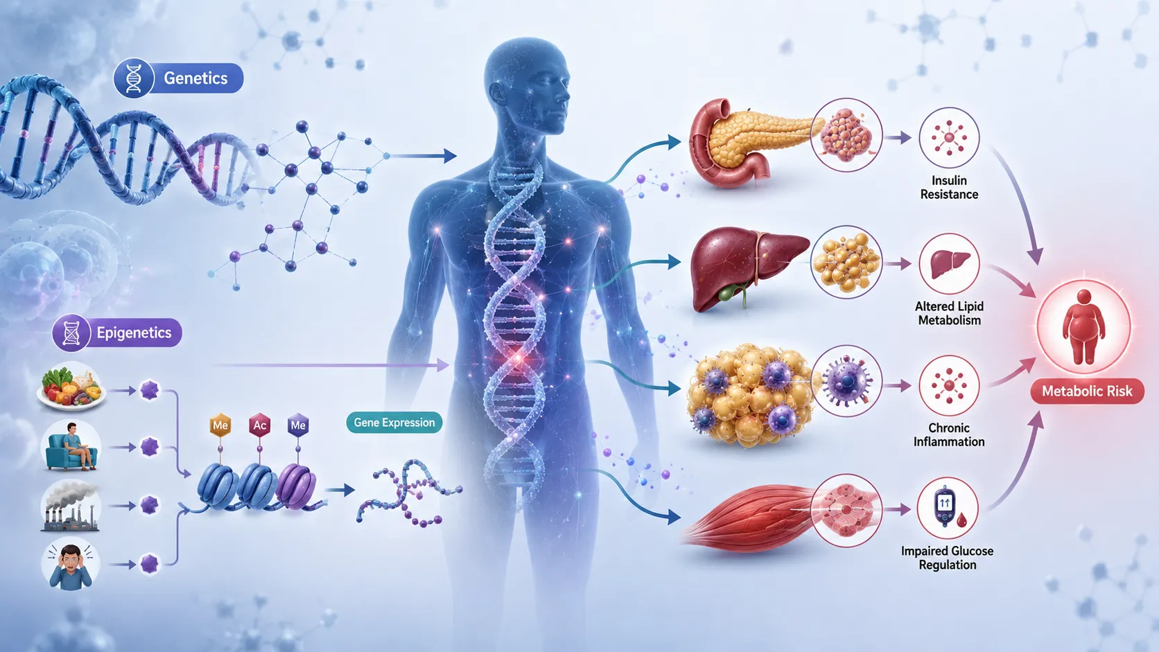
Key Definitions and Concepts
| Term | Definition | Relevance to Metabolic Disease |
|---|---|---|
| Genome | The complete set of genetic material contained within an organism. | Contains genes and variants that may influence metabolic susceptibility. |
| Gene | A functional DNA sequence that contributes to the production or regulation of biological molecules. | May influence insulin signalling, appetite, lipid metabolism and energy regulation. |
| Genetic variant | A difference in DNA sequence between individuals. | Some variants are associated with altered metabolic risk. |
| Single-nucleotide polymorphism (SNP) | A common variation involving a single DNA base. | May contribute small changes to metabolic disease susceptibility. |
| Epigenetics | Regulation of gene activity without changing the underlying DNA sequence. | Helps explain how environmental factors influence metabolic gene expression. |
| DNA methylation | Addition of methyl groups to DNA that can influence gene activity. | May alter the expression of genes involved in metabolism. |
| Histone modification | Chemical modification of proteins around which DNA is organised. | Can influence how accessible genes are for transcription. |
| Gene expression | The process through which genetic information is used to produce functional biological products. | Determines which metabolic pathways are active in specific tissues. |
| Polygenic risk | Combined influence of multiple genetic variants on disease susceptibility. | Explains why common metabolic diseases rarely depend on one gene. |
| Gene–environment interaction | A situation in which genetic effects vary according to environmental exposure. | Important in explaining different responses to diet and lifestyle. |
| Epigenetic plasticity | The capacity of epigenetic patterns to change in response to biological or environmental influences. | May contribute to adaptation or maladaptation in metabolic health. |
Understanding Biological Predisposition to Metabolic Disease
Biological predisposition refers to characteristics that increase the likelihood of developing a particular condition. It does not mean that the condition will inevitably occur.
Metabolic diseases are usually multifactorial. This means that their development involves multiple interacting influences.
Major Contributors May Include
Inherited genetic variation.
Epigenetic regulation.
Dietary patterns.
Physical activity.
Sleep and circadian patterns.
Age.
Hormonal status.
Adipose tissue distribution.
Environmental exposures.
Socioeconomic and behavioural influences.
Existing medical conditions.
Genetic and epigenetic mechanisms should therefore be considered within a broader systems-based model of metabolic health.
The Difference Between Genetics and Epigenetics
Genetics primarily concerns the inherited DNA sequence. A person inherits genetic material from their biological parents, and this genetic sequence provides instructions for many biological processes.
Epigenetics concerns mechanisms that influence how genetic information is used. Epigenetic processes can affect whether particular genes are more or less active in a specific tissue.
A Simplified Comparison
Genetics may influence:
The structure of a metabolic protein.
The efficiency of an enzyme.
The function of a receptor.
The biological tendency towards a particular metabolic response.
Epigenetics may influence:
Whether a gene is actively transcribed.
The degree of gene expression.
Tissue-specific patterns of gene activity.
The response of genes to environmental conditions.
Together, these systems contribute to individual metabolic diversity.
Genetic Architecture of Common Metabolic Diseases
Most common metabolic diseases are polygenic rather than caused by a single genetic variant.
A polygenic condition results from the combined effects of multiple genetic variants, each of which may contribute a relatively small effect to overall risk.
Polygenic Susceptibility Can Influence
Appetite regulation.
Food preference.
Energy balance.
Body fat distribution.
Insulin sensitivity.
Insulin secretion.
Hepatic metabolism.
Lipid transport.
Inflammatory responses.
The cumulative effect of many variants may create a higher or lower level of biological susceptibility.
Monogenic and Polygenic Metabolic Disorders
It is important to distinguish between monogenic and polygenic conditions.
Monogenic Disorders
Monogenic disorders result primarily from changes affecting a single gene.
Characteristics may include:
Stronger inheritance patterns.
Earlier clinical presentation in some conditions.
More clearly defined molecular mechanisms.
Potentially severe metabolic consequences.
Polygenic Disorders
Polygenic disorders involve multiple genetic variants.
Characteristics include:
Complex inheritance.
Strong interaction with environmental factors.
Variable clinical presentation.
Difficulties predicting individual outcomes.
Most common obesity and type 2 diabetes patterns involve polygenic and environmental interactions.
Genetic Factors Affecting Insulin Secretion
Genetic variation can influence pancreatic beta-cell development and function.
The ability of beta cells to respond to increased metabolic demand is an important factor in determining whether insulin resistance progresses towards persistent hyperglycaemia.
Genetic Influences May Affect
Beta-cell development.
Insulin synthesis.
Insulin processing.
Glucose sensing.
Insulin secretion.
Cellular resilience under metabolic stress.
Individuals with reduced beta-cell reserve may be less able to compensate for increasing insulin resistance.
Genetic Factors Affecting Insulin Sensitivity
Genetic variation may also influence how effectively tissues respond to insulin.
Important tissues include:
Skeletal muscle.
Liver.
Adipose tissue.
Genetic differences can influence proteins involved in:
Insulin receptor signalling.
Glucose transport.
Lipid oxidation.
Mitochondrial activity.
Adipocyte differentiation.
These differences may alter the metabolic response to nutrient excess.
Genetic Influences on Body Fat Distribution
Body fat distribution is an important determinant of metabolic risk.
Genetic factors can influence whether excess energy is more likely to be stored in:
Subcutaneous adipose tissue.
Visceral adipose tissue.
Ectopic locations such as the liver.
Individuals with similar body mass can therefore have different levels of metabolic risk.
Genetic Differences May Influence
Adipocyte number.
Adipocyte expansion capacity.
Fat storage location.
Lipid mobilisation.
Adipose tissue inflammation.
These factors help explain why body weight alone does not fully predict metabolic health.
Genetic Regulation of Appetite and Energy Balance
Energy balance is influenced by complex interactions between the brain, gastrointestinal system, adipose tissue and endocrine system.
Genetic variation may affect:
Hunger signalling.
Satiety responses.
Food reward pathways.
Energy expenditure.
Physical activity tendencies.
These biological influences interact with the food environment and behavioural patterns.
Genetic predisposition should not be interpreted as a lack of personal control. Biological susceptibility is only one component of a complex metabolic system.
Polygenic Risk Scores
A polygenic risk score combines information from multiple genetic variants to estimate an individual’s inherited susceptibility to a particular condition.
These tools are increasingly used in research.
Potential Advantages Include
Identification of population-level risk patterns.
Improved understanding of genetic architecture.
Investigation of gene–environment interactions.
Potential future support for personalised prevention.
Important Limitations Include
Predictive performance can vary between populations.
Risk scores may not capture environmental influences.
They do not provide certainty about disease development.
Ethical and privacy concerns must be considered.
Polygenic information should therefore be interpreted carefully.
Epigenetics and Metabolic Regulation
Epigenetic regulation helps determine which genes are active in particular cells.
Although most cells contain broadly the same DNA sequence, liver cells, muscle cells and adipocytes have different functions because they express different combinations of genes.
Epigenetic mechanisms contribute to this regulation.
Major Epigenetic Mechanisms Include
DNA methylation.
Histone modification.
Chromatin remodelling.
Regulation by non-coding RNA molecules.
These processes can influence metabolic pathways and cellular adaptation.
DNA Methylation
DNA methylation involves the addition of methyl groups to particular DNA regions.
The effect of methylation on gene activity depends on its location and biological context.
Changes in DNA Methylation May Be Associated With
Nutrient availability.
Ageing.
Inflammation.
Metabolic stress.
Environmental exposure.
Hormonal influences.
Altered methylation patterns have been investigated in relation to obesity, insulin resistance and type 2 diabetes.
However, association does not automatically demonstrate causation.
Histone Modifications
DNA is organised around proteins called histones. Chemical modifications to histones can influence how tightly DNA is packaged.
When DNA is more accessible, certain genes may be more readily transcribed. When access is restricted, gene activity may decrease.
Important Histone-Related Processes Include
Acetylation.
Methylation.
Other chemical modifications.
These mechanisms contribute to the regulation of metabolic gene expression.
Chromatin Remodelling
Chromatin refers to the complex structure formed by DNA and associated proteins.
Chromatin remodelling can alter the physical accessibility of specific genetic regions.
This allows cells to respond dynamically to internal and external signals.
Metabolic Influences on Chromatin Regulation May Include
Nutrient availability.
Hormonal signalling.
Cellular energy status.
Inflammation.
Oxidative stress.
Persistent metabolic stress may therefore influence long-term patterns of gene regulation.
Non-Coding RNA and Metabolic Disease
Not all RNA molecules are used to produce proteins. Some non-coding RNAs help regulate gene expression.
MicroRNAs and other regulatory RNA molecules have been investigated for their role in:
Insulin signalling.
Adipocyte development.
Lipid metabolism.
Inflammatory regulation.
Beta-cell function.
These mechanisms are an important area of emerging metabolic research.
Nutritional Influences on Epigenetic Regulation
Nutrition provides both energy and molecular substrates that influence cellular metabolism.
Some nutrients and dietary patterns may influence pathways involved in epigenetic regulation.
Potential Nutritional Influences Include
Availability of methyl-group donors.
Overall energy intake.
Macronutrient balance.
Micronutrient status.
Bioactive food compounds.
The relationship between diet and epigenetic change is complex and should not be oversimplified.
A single food or nutrient should not be assumed to permanently modify disease risk through epigenetic mechanisms.
Developmental Programming and Metabolic Risk
Early life is a period of substantial biological development. Environmental conditions during fetal development and early childhood may influence long-term metabolic health.
The developmental origins framework proposes that early biological adaptation may influence susceptibility to later disease.
Important Early-Life Influences May Include
Maternal nutritional status.
Maternal metabolic health.
Birth conditions.
Early growth patterns.
Childhood nutrition.
Epigenetic mechanisms are one possible biological pathway through which early environmental conditions may influence later metabolism.
Epigenetic Changes and Obesity
Obesity is associated with altered gene expression in several metabolically important tissues.
Research has investigated epigenetic differences in:
Adipose tissue.
Skeletal muscle.
Liver tissue.
Pancreatic cells.
These changes may influence metabolic processes such as:
Adipogenesis.
Lipid storage.
Inflammation.
Insulin signalling.
The relationship remains complex because obesity itself may cause epigenetic changes. Therefore, researchers must determine whether an epigenetic alteration is a cause, consequence or adaptive response.
Epigenetic Factors in Insulin Resistance
Epigenetic regulation may influence genes involved in insulin action and glucose metabolism.
Potential consequences include altered expression of pathways related to:
Glucose transport.
Insulin receptor signalling.
Mitochondrial metabolism.
Lipid oxidation.
Inflammatory activity.
Persistent metabolic stress may modify gene regulation and contribute to reduced metabolic flexibility.
The Interaction Between Genetics and Epigenetics
Genetics and epigenetics should not be viewed as completely separate systems.
Genetic variation can influence susceptibility to epigenetic modification, while epigenetic processes can influence how genetic instructions are expressed.
A Combined Interaction May Follow This Pattern
An individual inherits genetic variants associated with metabolic susceptibility.
Environmental conditions create metabolic stress.
Cellular signalling pathways respond to the environment.
Epigenetic regulation alters patterns of gene expression.
Metabolic pathways adapt or become dysregulated.
The risk of severe biochemical abnormalities may increase.
This interaction provides a more realistic explanation of individual metabolic variation.
Gene–Environment Interactions
The effects of genetic susceptibility may depend heavily on environmental exposure.
For example, a genetic tendency towards reduced insulin sensitivity may produce different outcomes depending on:
Dietary patterns.
Physical activity.
Sleep.
Body fat distribution.
Stress exposure.
Age.
This Means That
A genetic risk variant does not guarantee disease.
Environmental exposure does not affect every individual equally.
Preventive strategies may need to consider biological diversity.
Lifestyle interventions can remain valuable despite genetic susceptibility.
Gene–Diet Interactions
Gene–diet interactions describe situations in which genetic differences influence the metabolic response to dietary exposure.
Research has explored whether genetic variation affects responses to:
Dietary fat.
Carbohydrate intake.
Energy restriction.
Specific micronutrients.
Different dietary patterns.
Important Scientific Considerations
Evidence for personalised nutrition is developing, but not every proposed gene–diet relationship is sufficiently established for routine clinical application.
High-quality research should consider:
Replication.
Population diversity.
Study design.
Biological plausibility.
Magnitude of effect.
Epigenetic Responses to Physical Activity
Physical activity can influence metabolic gene expression and cellular signalling.
Research suggests that muscle tissue can demonstrate molecular adaptations associated with physical activity.
Potential effects include changes in pathways related to:
Glucose utilisation.
Mitochondrial function.
Lipid oxidation.
Insulin sensitivity.
This illustrates the concept of epigenetic and metabolic plasticity.
Severe Biochemical Dysregulation
Severe biochemical dysregulation refers to significant disturbances in the normal control of metabolic pathways.
In metabolic diseases, this may involve:
Persistent hyperglycaemia.
Marked insulin resistance.
Severe dyslipidaemia.
Excessive hepatic glucose production.
Ectopic lipid accumulation.
Significant inflammatory activity.
Impaired beta-cell function.
Genetic and epigenetic factors may influence how rapidly or severely these abnormalities develop.
Why Some Individuals Develop More Severe Disease
Individuals exposed to similar environmental conditions can experience different outcomes.
Potential explanations include:
Different polygenic risk profiles.
Variation in beta-cell reserve.
Differences in adipose tissue storage capacity.
Differences in body fat distribution.
Epigenetic responses to environmental stress.
Mitochondrial variation.
Different inflammatory responses.
These factors can influence the threshold at which metabolic compensation fails.
Practical Example: Two Individuals With Similar Lifestyle Exposure
Consider two adults with similar dietary patterns and low physical activity levels.
Individual A
May demonstrate:
Moderate weight gain.
Preserved glucose regulation.
Relatively effective insulin secretion.
Lower visceral fat accumulation.
Individual B
May demonstrate:
Increased visceral adiposity.
Early hyperinsulinaemia.
Greater dyslipidaemia.
Rapid deterioration in glucose tolerance.
The difference may reflect a combination of genetic susceptibility, tissue-specific metabolic responses and epigenetic adaptation.
This example demonstrates why metabolic disease cannot be explained by lifestyle factors alone.
Investigating Genetic Factors in Clinical and Research Settings
The investigation of genetic susceptibility should follow appropriate scientific and ethical procedures.
A Systematic Process May Include
Defining the metabolic phenotype.
Collecting relevant clinical and family history.
Identifying appropriate research or clinical indications.
Using validated genetic testing where justified.
Interpreting results within population and clinical context.
Considering environmental and lifestyle influences.
Providing appropriate professional guidance.
Genetic results should not be interpreted outside appropriate expertise.
Investigating Epigenetic Factors
Epigenetic research requires careful methodological design.
Researchers may investigate:
DNA methylation patterns.
Histone modifications.
Gene expression.
Regulatory RNA molecules.
Important Methodological Considerations Include
Tissue specificity.
Timing of sample collection.
Age and sex differences.
Environmental exposures.
Medication use.
Disease stage.
Laboratory methodology.
Epigenetic findings from one tissue may not accurately represent another tissue.
Tissue Specificity
One of the major challenges in metabolic epigenetic research is tissue specificity.
A blood sample may be relatively easy to obtain, but metabolic dysfunction may originate primarily in:
Liver tissue.
Skeletal muscle.
Visceral adipose tissue.
Pancreatic tissue.
Therefore, Researchers Must Consider
Whether the sampled tissue reflects the biological process of interest.
Whether blood-based markers act as useful proxies.
Whether tissue-specific findings can be generalised.
This is essential for critical evaluation of research findings.
Temporal Changes in Epigenetic Patterns
Epigenetic patterns are not always fixed throughout life.
They may change in response to:
Ageing.
Weight change.
Dietary modification.
Physical activity.
Disease progression.
Medical treatment.
This creates an important challenge: an observed epigenetic difference may represent a cause, consequence or response to metabolic disease.
Benefits of Investigating Genetic and Epigenetic Factors
Understanding these factors can provide several benefits.
Scientific Benefits
Improved understanding of disease mechanisms.
Identification of biological pathways.
Recognition of population differences.
Development of research hypotheses.
Clinical Benefits
Improved risk stratification in selected settings.
Potential identification of individuals requiring earlier monitoring.
Support for personalised approaches where evidence is sufficient.
Improved understanding of variable treatment responses.
Public Health Benefits
Recognition that metabolic risk is biologically heterogeneous.
Improved targeting of preventive research.
Better understanding of life-course influences.
Limitations of Genetic and Epigenetic Information
Genetic and epigenetic information must be interpreted cautiously.
Important Limitations Include
Genetic risk does not equal disease certainty.
Individual variants may have small effects.
Polygenic scores may perform differently across populations.
Epigenetic associations may not prove causation.
Tissue-specific findings may be difficult to obtain.
Environmental exposures are difficult to measure accurately.
Results may change as scientific evidence develops.
These limitations prevent overly deterministic interpretations.
Ethical Considerations
Genetic and epigenetic investigations involve important ethical responsibilities.
Key Considerations Include
Informed consent.
Data privacy.
Confidentiality.
Appropriate data storage.
Clear communication of uncertainty.
Avoidance of genetic discrimination.
Fair access to emerging technologies.
Professionals must avoid presenting genetic information in a way that causes unnecessary fear or falsely promises certainty.
Practical Application in Metabolic Disease Research
A research project investigating severe metabolic dysregulation could integrate multiple data streams.
Clinical Information
Body composition.
Waist measurements.
Blood pressure.
Family history.
Dietary information.
Physical activity patterns.
Biochemical Information
Fasting glucose.
Longer-term glucose markers.
Insulin-related measures.
Lipid profiles.
Markers relevant to metabolic and inflammatory status.
Molecular Information
Genetic variation.
Gene expression.
Epigenetic patterns where appropriate.
Combining these data may provide a more comprehensive picture of metabolic risk.
A Framework for Critical Investigation
Learners can use the following framework when evaluating genetic and epigenetic contributions.
Step 1: Define the Metabolic Problem
Identify whether the focus is:
Obesity.
Insulin resistance.
Type 2 diabetes.
Dyslipidaemia.
Metabolic syndrome.
Step 2: Identify the Biological Pathways
Consider pathways related to:
Insulin signalling.
Beta-cell function.
Lipid metabolism.
Adipose tissue biology.
Appetite regulation.
Inflammation.
Step 3: Evaluate Genetic Evidence
Ask:
Is the association replicated?
What is the effect size?
Does the evidence apply to the population?
Is there biological plausibility?
Step 4: Evaluate Epigenetic Evidence
Ask:
Which tissue was studied?
Was the design longitudinal or cross-sectional?
Could disease status influence the epigenetic finding?
Are environmental confounders adequately addressed?
Step 5: Integrate Environmental Factors
Consider:
Nutrition.
Physical activity.
Sleep.
Stress.
Medication.
Socioeconomic conditions.
Step 6: Draw a Balanced Conclusion
Avoid deterministic statements and recognise uncertainty.
Common Errors in Interpretation
Learners should avoid several common mistakes.
Error 1: Assuming a Gene Causes Disease Directly
Most common metabolic diseases involve multiple interacting genes and environmental factors.
Error 2: Treating Genetic Risk as a Prediction of Certainty
Risk is probabilistic rather than deterministic.
Error 3: Assuming Epigenetic Changes Are Always Permanent
Some epigenetic patterns can change in response to biological and environmental conditions.
Error 4: Confusing Association With Causation
An observed molecular difference may be a consequence rather than a cause of disease.
Error 5: Ignoring Population Diversity
Genetic findings may not have equal predictive value across all populations.
Error 6: Ignoring Environmental Influences
Genetic and epigenetic mechanisms operate within environmental contexts.
Professional Relevance for Learners
Understanding genetic and epigenetic mechanisms can strengthen professional competence in clinical nutrition and metabolic health.
Learners should be able to:
Explain the difference between genetic and epigenetic regulation.
Recognise the polygenic nature of common metabolic diseases.
Understand gene–environment interactions.
Interpret molecular evidence critically.
Recognise limitations and uncertainty.
Communicate biological risk without deterministic language.
Work within professional and ethical boundaries.
Key Learning Points
The following principles summarise the major concepts.
Genetics
Genetic variation contributes to individual metabolic susceptibility.
Common obesity and type 2 diabetes are generally polygenic.
Genes may influence insulin secretion and sensitivity.
Genetic variation can influence body fat distribution.
Genetic risk does not guarantee disease development.
Epigenetics
Epigenetic mechanisms regulate gene activity without changing DNA sequence.
DNA methylation and histone modification influence gene expression.
Environmental exposures may affect epigenetic regulation.
Epigenetic patterns can be tissue-specific and dynamic.
Epigenetic associations do not automatically establish causation.
Combined Effects
Genetics and epigenetics interact with environmental exposure.
Nutritional and lifestyle factors may influence molecular pathways.
Different individuals may respond differently to similar exposures.
Severe biochemical dysregulation may result when multiple risk factors interact.
Summary
The development of severe biochemical dysregulation in metabolic diseases is influenced by a complex combination of genetic susceptibility, epigenetic regulation and environmental exposure. Genetic variation can affect insulin secretion, insulin sensitivity, appetite regulation, body fat distribution and lipid metabolism. Most common metabolic diseases are polygenic, meaning that many genetic variants combine to influence overall susceptibility.
Epigenetic mechanisms add another layer of biological regulation by influencing how genes are expressed. DNA methylation, histone modifications, chromatin remodelling and non-coding RNA regulation can affect metabolic pathways without changing the underlying DNA sequence. These processes may respond to factors such as nutrition, physical activity, inflammation and metabolic stress.
The interaction between genetics and epigenetics helps explain why individuals exposed to similar lifestyle conditions can experience different metabolic outcomes. However, biological predisposition must never be interpreted as predetermined disease. Genetic risk is probabilistic, and environmental factors remain important.
A rigorous investigation of genetic and epigenetic influences requires critical analysis of molecular evidence, tissue specificity, study design and potential confounding factors. Learners must distinguish association from causation and recognise that epigenetic changes may be causes, consequences or adaptive responses to metabolic disease.
By understanding these combined mechanisms, Learners can develop a more sophisticated view of obesity, insulin resistance and metabolic syndrome. This knowledge supports evidence-based research, responsible interpretation of emerging molecular technologies and improved understanding of the biological diversity that characterises metabolic disease.
5.Synthesise Current Scientific Models to Explain the Active Role of Adipose Tissue as an Endocrine Organ Contributing to Metabolic Disease Pathology
Adipose tissue was traditionally regarded as a relatively passive storage site for excess energy in the form of triglycerides. Contemporary scientific research has fundamentally changed this understanding. Adipose tissue is now recognised as a highly active endocrine, metabolic and immunological organ that communicates continuously with the brain, liver, skeletal muscle, pancreas, immune system and cardiovascular system.
This communication occurs through the production and release of biologically active substances, including adipokines, cytokines, hormones, lipids and extracellular signalling molecules. Under healthy conditions, adipose tissue contributes to energy regulation, insulin sensitivity, appetite control, reproductive function and immune homeostasis. However, when adipose tissue expands excessively or becomes dysfunctional, these regulatory functions may become disrupted.
Dysfunctional adipose tissue is strongly associated with insulin resistance, chronic low-grade inflammation, dyslipidaemia, ectopic fat accumulation and disturbances in glucose metabolism. These interconnected abnormalities contribute significantly to the development and progression of obesity, type 2 diabetes, metabolic syndrome and cardiovascular disease.
Understanding adipose tissue as an endocrine organ is therefore essential for explaining the biochemical mechanisms underlying metabolic disease pathology.
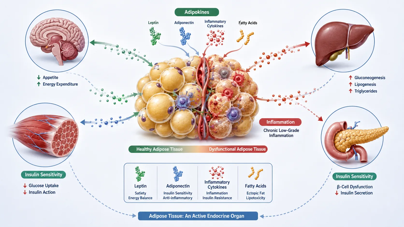
Key Definitions and Concepts
| Term | Definition | Clinical and Metabolic Significance |
|---|---|---|
| Adipose tissue | A specialised connective tissue composed primarily of adipocytes and supporting cells | Regulates energy storage and releases endocrine signalling molecules |
| Adipocyte | A specialised cell responsible for storing and mobilising lipids | Produces adipokines and participates in metabolic regulation |
| Endocrine organ | An organ that produces signalling molecules capable of influencing distant tissues | Adipose tissue influences multiple metabolic organs |
| Adipokine | A biologically active molecule produced or regulated by adipose tissue | Can influence inflammation, insulin sensitivity and energy balance |
| Leptin | An adipose-derived hormone involved in appetite and energy regulation | Dysregulation may contribute to leptin resistance and obesity |
| Adiponectin | An adipokine associated with improved insulin sensitivity | Reduced levels are associated with metabolic dysfunction |
| Chronic low-grade inflammation | Persistent activation of inflammatory pathways at relatively low intensity | Contributes to insulin resistance and metabolic disease |
| Lipotoxicity | Cellular dysfunction caused by excessive exposure to lipids or lipid metabolites | Damages metabolic tissues such as the liver and skeletal muscle |
| Ectopic fat | Fat accumulation in tissues not specialised for large-scale lipid storage | Associated with insulin resistance and organ dysfunction |
| Adipose tissue dysfunction | Impaired capacity of adipose tissue to safely store and regulate energy | A major contributor to metabolic disease progression |
The Evolution of Scientific Understanding of Adipose Tissue
The Traditional Energy Storage Model
Historically, adipose tissue was mainly viewed as a reservoir for excess energy. When energy intake exceeded energy expenditure, nutrients were converted into triglycerides and stored within adipocytes. During periods of fasting or increased energy demand, stored triglycerides were broken down through lipolysis, releasing fatty acids for use as an energy source.
This model remains important, but it is incomplete. Scientific advances have demonstrated that adipose tissue actively senses nutritional status and communicates this information throughout the body.
The traditional functions include:
Storage of triglycerides during periods of energy surplus.
Release of fatty acids during fasting or increased energy demand.
Physical protection and cushioning of organs.
Contribution to thermal insulation.
Participation in reproductive and developmental processes.
However, modern research demonstrates that adipose tissue also:
Produces hormones.
Releases inflammatory mediators.
Regulates insulin responsiveness.
Influences appetite and energy expenditure.
Communicates with the central nervous system.
Affects vascular function.
Interacts with immune cells.
Influences liver and skeletal muscle metabolism.
The Modern Endocrine Model
The endocrine model recognises adipose tissue as an active signalling system. Adipocytes and other cells within adipose tissue release numerous molecules that influence distant organs.
Adipose tissue contains more than mature adipocytes. Its cellular environment includes:
Preadipocytes.
Macrophages.
T lymphocytes.
B lymphocytes.
Endothelial cells.
Fibroblasts.
Vascular cells.
Other immune and stromal cells.
Together, these components form a complex biological environment often referred to as the adipose tissue microenvironment.
Changes within this environment can substantially alter metabolic health. For example, excessive adipose expansion may increase immune cell recruitment and inflammatory signalling, contributing to systemic metabolic dysfunction.
Types and Distribution of Adipose Tissue
White Adipose Tissue
White adipose tissue is the principal site of energy storage in humans. Its adipocytes contain large lipid droplets that store triglycerides.
White adipose tissue performs several important functions:
Stores excess energy.
Releases fatty acids when energy is required.
Produces leptin.
Produces adiponectin.
Releases inflammatory mediators.
Participates in endocrine communication.
White adipose tissue is not metabolically identical throughout the body. Its location significantly affects its physiological consequences.
Subcutaneous Adipose Tissue
Subcutaneous adipose tissue is located beneath the skin. It is generally considered to have a greater capacity for relatively safe lipid storage compared with excessive accumulation of fat around internal organs.
Important characteristics include:
Significant energy storage capacity.
Relatively lower exposure of the liver to released fatty acids.
Potential protective storage capacity when functioning normally.
Variable endocrine activity depending on metabolic health.
Healthy expansion of subcutaneous adipose tissue may reduce the need for lipid accumulation in other organs.
Visceral Adipose Tissue
Visceral adipose tissue surrounds internal organs within the abdominal cavity. Excessive accumulation is strongly associated with adverse metabolic outcomes.
Visceral adipose tissue is particularly important because:
It is metabolically active.
It can release inflammatory mediators.
Its products may influence liver metabolism.
It is associated with insulin resistance.
It is associated with an increased risk of metabolic syndrome.
The distinction between subcutaneous and visceral fat demonstrates that total body fat alone does not fully explain metabolic risk. Distribution and functional characteristics are also highly important.
Brown and Thermogenic Adipose Tissue
Brown adipose tissue has a specialised role in energy expenditure and heat production. It contains numerous mitochondria and can convert chemical energy into heat.
Thermogenic adipose activity is relevant because it demonstrates that adipose tissue can participate actively in energy expenditure rather than functioning solely as a storage compartment.
Key characteristics include:
High mitochondrial content.
Capacity for thermogenesis.
Participation in energy balance.
Potential influence on glucose and lipid metabolism.
Current scientific models continue to investigate whether altered thermogenic adipose activity contributes to metabolic disease risk.
Adipose Tissue as an Endocrine Signalling Organ
Adipokines and Systemic Communication
Adipokines are signalling molecules produced or regulated by adipose tissue. They influence numerous physiological processes.
The effects of adipokines may include:
Regulation of appetite.
Control of insulin sensitivity.
Modulation of inflammation.
Regulation of energy expenditure.
Influence on vascular function.
Effects on lipid metabolism.
The overall metabolic effect depends on the balance of different signalling molecules rather than the action of a single hormone.
Leptin and Energy Regulation
Leptin is produced primarily by adipose tissue and communicates information about energy stores to the central nervous system. Under normal physiological conditions, leptin contributes to appetite regulation and energy balance.
A simplified physiological sequence is:
Adipose tissue energy stores increase.
Leptin production generally increases.
Leptin signals the central nervous system.
Appetite and energy expenditure pathways respond.
Energy balance is regulated.
In obesity, circulating leptin concentrations may be elevated while physiological responsiveness is reduced. This phenomenon is commonly described as leptin resistance.
Potential consequences include:
Reduced effectiveness of appetite regulation.
Continued excessive energy intake.
Altered neuroendocrine signalling.
Further adipose tissue expansion.
Leptin resistance illustrates an important principle of endocrine pathology: higher concentrations of a signalling molecule do not necessarily produce a stronger biological effect.
Adiponectin and Insulin Sensitivity
Adiponectin is an adipokine strongly associated with favourable metabolic regulation. Unlike some adipose-derived signals, adiponectin levels may decrease as adipose tissue dysfunction and metabolic disease become more severe.
Adiponectin is associated with:
Improved insulin sensitivity.
Enhanced fatty acid metabolism.
Reduced metabolic stress.
Potential anti-inflammatory effects.
Improved metabolic communication between tissues.
Reduced adiponectin may therefore contribute to:
Insulin resistance.
Impaired lipid metabolism.
Increased metabolic inflammation.
Progression towards type 2 diabetes.
The relationship between adiponectin and obesity demonstrates that adipose tissue function is more important than simply the quantity of adipose tissue.
Adipose Tissue and Chronic Inflammation
The Healthy-to-Dysfunctional Transition
As adipose tissue expands, adipocytes may undergo substantial physiological stress. Excessive enlargement can disrupt oxygen supply, mechanical structure and metabolic regulation.
The progression may involve:
Adipocyte enlargement.
Local tissue stress.
Reduced oxygen availability in some regions.
Cellular stress signalling.
Recruitment of immune cells.
Increased inflammatory mediator production.
Progressive impairment of insulin signalling.
This creates an environment in which metabolic and immune processes become increasingly interconnected.
Immune Cell Infiltration
Adipose tissue contains immune cells under normal conditions. However, obesity-related adipose dysfunction can alter the number and activity of these cells.
Important cellular changes may include:
Increased macrophage accumulation.
Changes in macrophage functional states.
Increased inflammatory signalling.
Altered T-cell activity.
Greater cytokine production.
Macrophages can release inflammatory mediators that interfere with normal metabolic signalling.
This illustrates why obesity is increasingly understood as involving both metabolic and immunological dysregulation.
Pro-Inflammatory Cytokines
Dysfunctional adipose tissue can contribute to increased production of inflammatory mediators, including cytokines that influence insulin-sensitive tissues.
Persistent inflammatory signalling may contribute to:
Impaired insulin receptor signalling.
Altered glucose uptake.
Increased hepatic glucose production.
Disturbed lipid metabolism.
Endothelial dysfunction.
The important clinical principle is that chronic metabolic inflammation differs from an acute protective immune response. It may persist over long periods and contribute progressively to tissue dysfunction.
Molecular Models of Insulin Resistance
Normal Insulin Signalling
Insulin regulates glucose and nutrient metabolism in several tissues.
Under normal conditions:
Insulin binds to its receptor.
Intracellular signalling pathways become activated.
Glucose transport mechanisms respond.
Skeletal muscle and adipose tissue increase glucose uptake.
The liver reduces inappropriate glucose production.
Blood glucose levels remain within an appropriate physiological range.
This coordinated process depends on effective communication between multiple organs.
Disruption by Inflammatory Signalling
Chronic inflammatory signals can interfere with components of insulin signalling pathways.
Potential mechanisms include:
Altered phosphorylation of signalling proteins.
Cellular stress responses.
Activation of inflammatory kinase pathways.
Changes in mitochondrial function.
Accumulation of harmful lipid intermediates.
The outcome may be reduced cellular responsiveness to insulin.
As insulin resistance progresses:
The pancreas may initially increase insulin secretion.
Blood insulin levels may rise.
Glucose control may remain temporarily compensated.
Beta-cell stress may increase over time.
Glucose concentrations may eventually rise.
This progression helps explain the development of type 2 diabetes.
Adipose Tissue Lipolysis and Metabolic Disease
Normal Lipid Mobilisation
Lipolysis is the breakdown of stored triglycerides into fatty acids and glycerol.
It is an essential physiological process during:
Fasting.
Exercise.
Increased energy demand.
Reduced nutrient availability.
The process must be tightly regulated.
Dysregulated Lipolysis
In insulin-resistant adipose tissue, insulin may become less effective at suppressing lipolysis. This can increase the release of fatty acids into the circulation.
Excessive fatty acid availability may contribute to:
Increased liver lipid synthesis.
Elevated triglyceride concentrations.
Ectopic fat accumulation.
Skeletal muscle insulin resistance.
Pancreatic stress.
The relationship can create a self-reinforcing metabolic cycle.
The Metabolic Spillover Model
The metabolic spillover model proposes that when adipose tissue can no longer safely manage excess lipid storage, lipids increasingly accumulate in other tissues.
The sequence may involve:
Chronic energy surplus.
Adipocyte enlargement.
Reduced adipose storage flexibility.
Increased fatty acid release.
Lipid deposition in the liver.
Lipid accumulation in skeletal muscle.
Development of cellular metabolic stress.
Progressive insulin resistance.
This model explains why metabolic disease may develop when lipid storage becomes dysfunctional rather than simply because total fat mass increases.
Ectopic Fat Accumulation
Hepatic Fat Accumulation
The liver plays a central role in glucose and lipid metabolism. Excess fatty acid delivery can contribute to abnormal hepatic lipid accumulation.
Potential consequences include:
Increased triglyceride synthesis.
Altered lipoprotein production.
Hepatic insulin resistance.
Increased glucose production.
Further dyslipidaemia.
A metabolically dysfunctional liver can therefore contribute to systemic metabolic disease.
Skeletal Muscle Lipid Accumulation
Skeletal muscle is one of the body’s major sites of insulin-stimulated glucose uptake.
Excess intracellular lipid accumulation may interfere with normal metabolic signalling.
Potential consequences include:
Reduced glucose uptake.
Impaired mitochondrial metabolism.
Altered insulin signalling.
Reduced metabolic flexibility.
This demonstrates that insulin resistance is not restricted to adipose tissue.
Adipose Tissue and the Metabolic Syndrome
Understanding Metabolic Syndrome
Metabolic syndrome describes a cluster of interconnected metabolic abnormalities that increase the risk of major chronic disease.
Common components include:
Central adiposity.
Elevated blood glucose.
Insulin resistance.
Dyslipidaemia.
Elevated blood pressure.
Adipose tissue dysfunction provides a biological framework that helps connect these abnormalities.
The Multi-Organ Interaction Model
Modern scientific models emphasise that metabolic disease involves communication between organs.
The principal interacting systems include:
Adipose tissue.
Liver.
Skeletal muscle.
Pancreas.
Brain.
Immune system.
Cardiovascular system.
For example:
Dysfunctional adipose tissue releases excess fatty acids.
The liver develops metabolic disturbances.
Skeletal muscle becomes less responsive to insulin.
The pancreas increases insulin secretion.
Chronic inflammation intensifies.
Cardiovascular risk increases.
This multi-organ perspective is essential for understanding complex metabolic disease.
Adipose Tissue Hypoxia and Cellular Stress
Why Hypoxia May Occur
Rapid adipose tissue expansion can challenge the existing vascular supply. Some regions may experience reduced oxygen availability.
Reduced oxygen availability can contribute to:
Activation of stress-responsive pathways.
Altered mitochondrial function.
Increased inflammatory signalling.
Changes in extracellular matrix structure.
The precise effects may vary between individuals and adipose depots.
Endoplasmic Reticulum Stress
The endoplasmic reticulum is involved in protein processing and cellular homeostasis. Excess nutrient availability and metabolic stress can disrupt its normal function.
Potential consequences include:
Activation of stress pathways.
Altered inflammatory signalling.
Impaired insulin responsiveness.
Increased cellular dysfunction.
This demonstrates that metabolic disease involves processes occurring at both organ and cellular levels.
Adipose Tissue Remodelling
Healthy Adipose Expansion
Adipose tissue expansion is not always harmful. Under certain conditions, tissue can expand while maintaining appropriate blood supply, cellular function and metabolic regulation.
Healthier expansion may involve:
Formation of new adipocytes.
Adequate vascular adaptation.
Controlled extracellular matrix remodelling.
Limited inflammatory activation.
Continued insulin responsiveness.
Pathological Adipose Expansion
Pathological expansion may involve excessive enlargement of existing adipocytes rather than effective tissue adaptation.
Potential features include:
Adipocyte hypertrophy.
Tissue hypoxia.
Fibrosis.
Inflammation.
Reduced insulin sensitivity.
Impaired lipid storage capacity.
The distinction between hypertrophy and healthier tissue expansion is important when evaluating metabolic risk.
Key Scientific Models Explaining Adipose Tissue Pathology
The Adipose Tissue Expandability Model
This model proposes that metabolic health depends partly on the capacity of adipose tissue to safely store excess energy.
When storage capacity is exceeded:
Lipids may escape into the circulation.
Ectopic fat accumulation may increase.
Lipotoxicity may develop.
Insulin resistance may worsen.
The model helps explain why individuals with similar body mass may experience different metabolic outcomes.
The Inflammatory Model
The inflammatory model emphasises persistent immune activation within dysfunctional adipose tissue.
The proposed pathway includes:
Nutrient excess.
Adipocyte stress.
Release of distress signals.
Recruitment and activation of immune cells.
Cytokine production.
Interference with metabolic signalling.
Systemic insulin resistance.
A major benefit of this model is that it integrates metabolic and immune mechanisms.
The Adipokine Imbalance Model
This model focuses on changes in the balance between beneficial and harmful adipose-derived signals.
Potential changes include:
Reduced adiponectin.
Increased inflammatory mediators.
Altered leptin responsiveness.
Disturbed endocrine communication.
The resulting imbalance can influence multiple organs simultaneously.
The Ectopic Fat Model
This model focuses on the consequences of lipid accumulation outside normal adipose storage compartments.
Important target tissues include:
Liver.
Skeletal muscle.
Pancreas.
Heart.
Excess lipid exposure can contribute to cellular dysfunction and impaired metabolic signalling.
Practical Clinical Interpretation
Why Body Weight Alone Is Insufficient
Body weight and body mass index provide useful information but do not fully describe metabolic function.
Two individuals with similar body size may have different:
Fat distribution.
Visceral adiposity.
Insulin sensitivity.
Inflammatory activity.
Lipid profiles.
Liver fat accumulation.
Clinical assessment should therefore consider broader metabolic information.
Relevant Clinical Indicators
A comprehensive metabolic assessment may consider:
Waist circumference.
Blood glucose measures.
Indicators of long-term glycaemic control.
Lipid profile.
Blood pressure.
Liver-related biochemical findings.
Markers of systemic inflammation where clinically appropriate.
Clinical history.
Dietary patterns.
Physical activity.
Medication use.
No single measurement should be interpreted as a complete representation of adipose tissue health.
Practical Example: Progressive Adipose Tissue Dysfunction
Consider an adult who experiences prolonged energy surplus and gradual central weight gain.
Initially:
Adipose tissue stores increasing amounts of triglycerides.
Blood glucose remains relatively controlled.
Insulin secretion increases to maintain normal glucose regulation.
Over time:
Adipocytes enlarge.
Insulin responsiveness decreases.
Lipolysis becomes less effectively regulated.
Circulating fatty acid exposure increases.
Liver lipid accumulation develops.
Inflammatory signalling becomes more prominent.
Later stages may involve:
Increased fasting glucose.
Dyslipidaemia.
Reduced insulin sensitivity.
Increased risk of type 2 diabetes.
This example demonstrates how adipose tissue dysfunction can contribute progressively to multi-organ metabolic disease.
Benefits of Understanding Adipose Tissue as an Endocrine Organ
A modern understanding of adipose biology provides important benefits for clinical and scientific practice.
Improved Understanding of Disease Mechanisms
It helps Learners and healthcare professionals understand:
Why obesity is biologically complex.
How inflammation and metabolism interact.
Why insulin resistance involves multiple organs.
Why fat distribution influences metabolic risk.
More Comprehensive Clinical Assessment
The endocrine model encourages assessment beyond body weight alone.
This supports consideration of:
Metabolic markers.
Fat distribution.
Insulin sensitivity.
Lipid metabolism.
Inflammatory status.
Organ function.
Improved Therapeutic Development
Understanding molecular pathways may support the development of interventions targeting:
Insulin sensitivity.
Adipose inflammation.
Lipid metabolism.
Appetite regulation.
Energy expenditure.
Better Personalisation of Metabolic Care
Different individuals may experience metabolic disease through different combinations of mechanisms.
Potential differences include:
Predominantly hepatic insulin resistance.
Greater visceral adiposity.
Stronger inflammatory activity.
Reduced adipose storage capacity.
Significant ectopic lipid accumulation.
Understanding these differences supports a more individualised approach to metabolic assessment and management.
Limitations and Critical Considerations
Adipokines Should Not Be Interpreted in Isolation
Although adipokines are biologically important, a single measurement may not provide a complete clinical picture.
Interpretation may be influenced by:
Body composition.
Acute illness.
Chronic inflammation.
Medication.
Age.
Sex.
Disease state.
Association Does Not Always Establish Causation
Research may demonstrate that a particular adipokine is associated with metabolic disease without proving that it directly causes the condition.
Critical evaluation should consider:
Study design.
Biological plausibility.
Confounding factors.
Reproducibility.
Experimental evidence.
Metabolic Disease Is Multifactorial
Adipose dysfunction is important but does not operate independently.
Metabolic disease may also be influenced by:
Genetic factors.
Dietary patterns.
Physical inactivity.
Sleep disruption.
Environmental exposures.
Medication.
Ageing.
Hormonal factors.
A comprehensive scientific model must integrate these influences.
A Systematic Process for Synthesising Scientific Models
Step 1: Identify the Primary Metabolic Disturbance
Determine the major biological problem, such as:
Insulin resistance.
Excess lipid accumulation.
Chronic inflammation.
Impaired energy regulation.
Step 2: Identify the Role of Adipose Tissue
Consider whether adipose tissue contributes through:
Excess fatty acid release.
Altered adipokine production.
Immune activation.
Reduced storage capacity.
Abnormal endocrine signalling.
Step 3: Identify the Affected Organs
Evaluate interactions with:
Liver.
Skeletal muscle.
Pancreas.
Brain.
Cardiovascular system.
Step 4: Connect the Molecular and Clinical Findings
The Learner should connect:
Cellular signalling.
Tissue dysfunction.
Biochemical abnormalities.
Clinical symptoms.
Long-term disease outcomes.
Step 5: Evaluate Alternative Explanations
A scientifically rigorous interpretation should also consider:
Genetic predisposition.
Lifestyle factors.
Medication effects.
Coexisting disease.
Environmental influences.
Key Points for Professional Practice
When applying these concepts, Learners should remember that:
Adipose tissue is an active endocrine and metabolic organ.
Healthy adipose tissue helps regulate energy and metabolic homeostasis.
Excessive or dysfunctional adipose expansion can disrupt endocrine signalling.
Adipokine imbalance can influence insulin sensitivity and inflammation.
Chronic low-grade inflammation connects immune dysfunction with metabolic disease.
Excess fatty acid release can contribute to ectopic lipid accumulation.
Visceral adiposity is particularly associated with adverse metabolic outcomes.
Insulin resistance develops through complex interactions across multiple tissues.
Body weight alone cannot fully describe metabolic health.
Metabolic disease should be understood using an integrated multi-organ model.
Scientific models must be critically evaluated because no single mechanism explains every case.
Conclusion
Contemporary scientific models clearly demonstrate that adipose tissue is far more than a passive energy storage compartment. It functions as a dynamic endocrine, metabolic and immunological organ that continuously communicates with multiple physiological systems. Through the release of adipokines, fatty acids, cytokines and other signalling molecules, adipose tissue influences appetite regulation, energy balance, glucose metabolism, insulin sensitivity and inflammatory activity.
When adipose tissue remains metabolically healthy, it can safely store energy and support physiological homeostasis. However, excessive or dysfunctional expansion may lead to adipocyte stress, chronic low-grade inflammation, altered adipokine secretion, dysregulated lipolysis and reduced capacity for safe lipid storage. These disturbances can promote ectopic fat accumulation and progressive insulin resistance across the liver, skeletal muscle and other metabolically important tissues.
The adipose tissue expandability model, inflammatory model, adipokine imbalance model and ectopic fat model each provide valuable perspectives for understanding metabolic disease. When synthesised, these models demonstrate that obesity and related metabolic disorders are multi-system conditions involving complex interactions between endocrine, immune and metabolic pathways.
For clinical and scientific practice, the most effective approach is to assess adipose tissue dysfunction within a broader metabolic context rather than relying solely on body weight or a single biochemical marker. This integrated understanding provides a stronger foundation for analysing metabolic disease pathology, interpreting clinical findings and developing evidence-informed strategies for prevention and management.
6.Assess the Severe Biochemical Consequences of Altered Mitochondrial Function and Sustained Oxidative Stress on Long-Term Metabolic Health
Mitochondria are essential intracellular organelles responsible for producing much of the energy required to sustain human life. They convert nutrients derived from carbohydrates, fats and proteins into adenosine triphosphate (ATP), the principal energy currency used by cells. However, mitochondria perform functions that extend far beyond energy production. They regulate cellular signalling, calcium balance, reactive oxygen species production, apoptosis, intermediary metabolism and communication between metabolic tissues.
Altered mitochondrial function can therefore have widespread consequences for metabolic health. When mitochondrial energy production becomes inefficient, cells may experience an imbalance between energy demand and energy availability. At the same time, dysfunctional mitochondria may generate excessive reactive oxygen species (ROS), contributing to oxidative stress. Sustained oxidative stress can damage lipids, proteins, DNA and cellular structures, further impairing metabolic regulation.
The interaction between mitochondrial dysfunction and oxidative stress is particularly important in the development and progression of metabolic disorders. These processes are associated with insulin resistance, impaired glucose regulation, abnormal lipid metabolism, obesity-related complications, fatty liver disease and progressive cardiovascular and metabolic risk.
This section examines the biochemical mechanisms through which altered mitochondrial function and persistent oxidative stress influence long-term metabolic health. Learners should develop the ability to assess these interconnected processes using biochemical principles, laboratory findings and clinical reasoning.
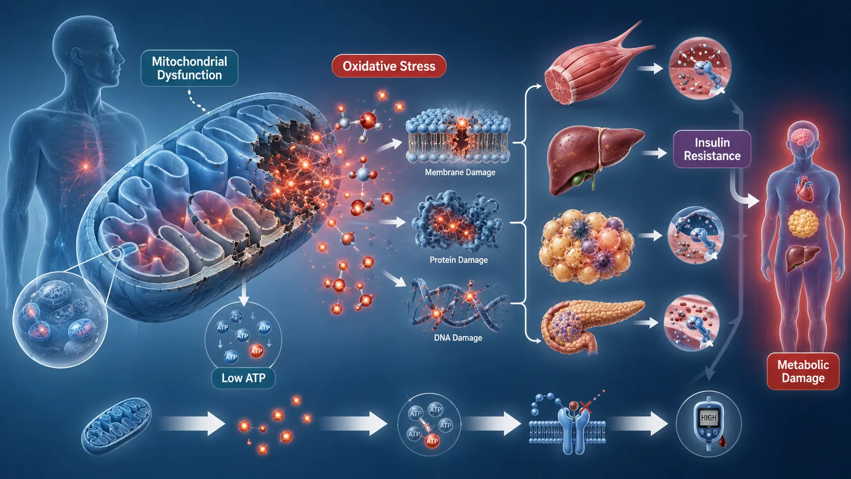
Key Concepts and Definitions
| Term | Definition | Relevance to Metabolic Health |
|---|---|---|
| Mitochondria | Cellular organelles that generate ATP and regulate multiple metabolic processes | Support energy production and metabolic flexibility |
| Oxidative phosphorylation | Process through which ATP is generated using the electron transport chain | Essential for efficient cellular energy production |
| Reactive oxygen species (ROS) | Chemically reactive oxygen-containing molecules produced during metabolism | Useful in signalling but harmful in excess |
| Oxidative stress | A state in which oxidant production exceeds antioxidant defence capacity | Can damage cellular components and disrupt metabolism |
| Mitochondrial dysfunction | Reduced or abnormal mitochondrial capacity to perform metabolic functions | Contributes to impaired glucose and lipid metabolism |
| Antioxidant defence | Enzymatic and non-enzymatic systems that neutralise excessive oxidants | Protects cells from oxidative damage |
| Metabolic flexibility | Ability to adapt fuel utilisation according to nutrient availability and energy demand | Important for glucose and lipid homeostasis |
| Electron transport chain | A series of mitochondrial protein complexes involved in energy generation | Dysfunction can reduce ATP and increase electron leakage |
| Lipid peroxidation | Oxidative damage to membrane lipids | Alters membrane function and cellular signalling |
| Mitochondrial biogenesis | Formation of new mitochondria within cells | Supports adaptation to changing metabolic demands |
Understanding the Central Role of Mitochondria in Metabolism
Mitochondria as Metabolic Control Centres
Mitochondria are often described as the energy-producing structures of the cell. Although this description is accurate, it is incomplete. Mitochria also act as metabolic control centres that coordinate nutrient oxidation, energy sensing and cellular adaptation.
Different tissues have different mitochondrial requirements. Skeletal muscle requires substantial energy during physical activity. The liver uses mitochondrial pathways to regulate nutrient processing and energy balance. Adipose tissue requires mitochondria for lipid metabolism and thermogenic processes, while pancreatic beta cells depend on mitochondrial metabolism to support appropriate insulin secretion.
Important mitochondrial functions include:
- ATP production through oxidative phosphorylation.
- Oxidation of fatty acids.
- Participation in carbohydrate metabolism.
- Processing of amino acid-derived metabolites.
- Regulation of calcium signalling.
- Generation and regulation of ROS.
- Control of programmed cell death.
- Support for cellular adaptation to energy demand.
- Regulation of intermediary metabolic pathways.
- Communication with the nucleus through metabolic signalling.
When these functions become impaired, metabolic disturbances can emerge across multiple organs.
ATP Production and Oxidative Phosphorylation
ATP production depends heavily on the electron transport chain, located within the inner mitochondrial membrane. Electrons derived from nutrient metabolism pass through a series of protein complexes. The energy released is used to establish a proton gradient, which drives ATP synthesis.
The simplified process involves:
- Nutrients are broken down through metabolic pathways.
- Electron carriers such as NADH and FADH₂ transfer high-energy electrons.
- Electrons enter the mitochondrial electron transport chain.
- Energy released during electron transfer contributes to proton movement.
- A proton gradient develops across the inner mitochondrial membrane.
- ATP synthase uses this gradient to generate ATP.
- ATP becomes available for cellular processes.
Efficient mitochondrial function is therefore essential for matching energy production with cellular demand.
The Biochemical Basis of Mitochondrial Dysfunction
What Is Mitochondrial Dysfunction?
Mitochondrial dysfunction refers to a reduction or alteration in normal mitochondrial capacity. It does not always mean that mitochondria have completely stopped functioning. Instead, mitochondrial efficiency, number, structure, signalling capacity or substrate utilisation may be impaired.
Mitochondrial dysfunction may involve:
- Reduced oxidative phosphorylation capacity.
- Impaired electron transport.
- Excessive electron leakage.
- Reduced ATP production.
- Abnormal mitochondrial dynamics.
- Impaired fatty acid oxidation.
- Altered mitochondrial DNA integrity.
- Reduced mitochondrial biogenesis.
- Disturbed calcium regulation.
- Excessive production of reactive oxygen species.
The consequences depend on the severity of dysfunction, the affected tissue and the individual’s overall metabolic state.
Causes of Altered Mitochondrial Function
Mitochondrial impairment can develop through multiple interacting influences rather than a single cause.
Potential contributing factors include:
- Chronic nutrient excess.
- Persistent hyperglycaemia.
- Excessive circulating fatty acids.
- Physical inactivity.
- Chronic inflammation.
- Genetic susceptibility.
- Age-related changes.
- Environmental stressors.
- Certain toxic exposures.
- Repeated cellular stress.
- Impaired antioxidant capacity.
A major principle in metabolic disease is that prolonged exposure to nutrient overload can exceed the capacity of cells to process and store metabolic substrates efficiently.
Nutrient Overload and Mitochondrial Stress
When excessive amounts of glucose and fatty acids repeatedly enter metabolic pathways, mitochondria may be exposed to increased substrate flux. Initially, cells may adapt by increasing energy production.
However, prolonged metabolic overload can contribute to:
- Increased electron transport activity.
- Greater electron leakage.
- Increased ROS production.
- Accumulation of incomplete metabolic intermediates.
- Lipid-derived cellular stress.
- Reduced metabolic flexibility.
- Progressive impairment of insulin signalling.
This demonstrates that nutrient availability is not always beneficial when it persistently exceeds metabolic requirements.
Reactive Oxygen Species and Their Physiological Role
Understanding Reactive Oxygen Species
Reactive oxygen species are produced naturally during normal metabolism. Common ROS-related molecules include superoxide and hydrogen peroxide. At controlled concentrations, these molecules contribute to normal cellular signalling.
Therefore, ROS should not be considered exclusively harmful.
Physiological functions of controlled ROS signalling include:
- Regulation of cellular adaptation.
- Participation in immune defence.
- Modulation of signalling pathways.
- Support for responses to exercise.
- Communication between cellular compartments.
- Regulation of gene expression.
Problems arise when ROS production becomes excessive or antioxidant defences become insufficient.
From Physiological Signalling to Oxidative Stress
Oxidative stress develops when there is a persistent imbalance between oxidant production and antioxidant protection.
The process can be understood as:
Normal metabolism → Controlled ROS production → Antioxidant neutralisation → Cellular balance
Under chronic stress:
Metabolic overload → Excess ROS generation → Antioxidant capacity overwhelmed → Molecular damage
Sustained oxidative stress can affect numerous cellular structures simultaneously.
Potential targets include:
- Cell membrane lipids.
- Structural proteins.
- Enzymes.
- Nuclear DNA.
- Mitochondrial DNA.
- Insulin signalling proteins.
- Components of the electron transport chain.
The Vicious Cycle Between Mitochondrial Dysfunction and Oxidative Stress
A Self-Reinforcing Biochemical Cycle
One of the most important concepts in metabolic disease is the reciprocal relationship between mitochondrial dysfunction and oxidative stress.
Mitochondrial dysfunction can increase ROS production. Excessive ROS can then damage mitochondrial structures, further worsening mitochondrial function.
This creates a progressive cycle:
- Mitochondrial efficiency declines.
- Electron transport becomes less efficient.
- Electron leakage increases.
- ROS production rises.
- Oxidative damage affects mitochondrial proteins and membranes.
- ATP production becomes further impaired.
- Metabolic stress increases.
- Additional ROS are generated.
Over time, this cycle can contribute to chronic metabolic dysfunction.
Why Mitochondrial DNA Is Vulnerable
Mitochondria contain their own genetic material, known as mitochondrial DNA. Because mitochondrial DNA is closely associated with the site of ROS production, it may be vulnerable to oxidative damage.
Potential consequences include:
- Impaired production of mitochondrial proteins.
- Reduced electron transport efficiency.
- Declining energy production.
- Increased susceptibility to further dysfunction.
- Reduced capacity for metabolic adaptation.
This can contribute to long-term changes in cellular energy metabolism.
Effects on Glucose Metabolism
Mitochondrial Dysfunction and Insulin Resistance
Insulin resistance occurs when target tissues show a reduced metabolic response to insulin. Skeletal muscle, liver and adipose tissue are particularly important in this process.
Mitochondrial dysfunction can contribute indirectly and directly to impaired insulin action. When nutrient oxidation is inefficient, lipid-derived metabolites may accumulate within cells.
These metabolic disturbances can interfere with signalling pathways involved in glucose uptake and utilisation.
Potential mechanisms include:
- Reduced metabolic flexibility.
- Incomplete fatty acid oxidation.
- Accumulation of lipid intermediates.
- Increased oxidative stress.
- Activation of stress-related signalling pathways.
- Impaired insulin signalling.
- Reduced glucose uptake in skeletal muscle.
The relationship is complex and should not be interpreted as a simple one-directional pathway. Insulin resistance itself may also worsen mitochondrial stress.
Skeletal Muscle and Glucose Disposal
Skeletal muscle is a major site of insulin-mediated glucose uptake. During healthy metabolic conditions, insulin promotes glucose transport into muscle cells, where glucose can be oxidised or stored.
Mitochondrial impairment may reduce the ability of muscle to adapt efficiently between carbohydrate and lipid oxidation.
This may result in:
- Reduced metabolic flexibility.
- Accumulation of metabolic intermediates.
- Reduced oxidative capacity.
- Greater susceptibility to lipid-induced insulin resistance.
- Impaired energy efficiency during physical activity.
Clinical Example
Consider an individual with obesity and gradually increasing fasting glucose concentrations. Laboratory assessment may eventually demonstrate:
- Elevated fasting glucose.
- Increased glycated haemoglobin.
- Elevated triglycerides.
- Reduced HDL cholesterol.
- Increased markers associated with metabolic dysfunction.
These findings do not directly measure mitochondrial function. However, the overall metabolic pattern may be consistent with processes in which impaired cellular energy regulation, insulin resistance and oxidative stress contribute to disease progression.
Effects on Lipid Metabolism
Fatty Acid Oxidation
Mitochondria play an essential role in fatty acid oxidation. Fatty acids must be transported and processed through several biochemical steps before they can contribute to energy production.
When mitochondrial fatty acid oxidation is impaired, cells may have difficulty processing lipid substrates efficiently.
Potential consequences include:
- Accumulation of lipid intermediates.
- Increased intracellular lipid storage.
- Reduced metabolic flexibility.
- Cellular stress.
- Impaired insulin sensitivity.
Lipotoxicity
Lipotoxicity refers to cellular dysfunction caused by excessive accumulation of lipid-related molecules in tissues not specialised for long-term fat storage.
For example, excessive lipid accumulation in:
- Skeletal muscle.
- Liver.
- Pancreatic tissue.
- Cardiac tissue.
may interfere with normal cellular function.
Lipotoxic stress can contribute to:
- Mitochondrial impairment.
- Endoplasmic reticulum stress.
- Oxidative stress.
- Inflammatory signalling.
- Insulin resistance.
- Cellular injury.
Mitochondrial Dysfunction in the Liver
The liver plays a central role in carbohydrate and lipid metabolism. Hepatic mitochondrial dysfunction can influence the handling of fatty acids and contribute to abnormal lipid accumulation.
Long-term disturbances may involve:
- Increased hepatic lipid storage.
- Altered fatty acid oxidation.
- Increased oxidative stress.
- Impaired glucose regulation.
- Progressive metabolic dysfunction.
The clinical significance of these processes must always be interpreted alongside the individual’s broader health status and diagnostic findings.
Oxidative Damage to Lipids, Proteins and DNA
Lipid Peroxidation
Cell membranes contain lipids that can be vulnerable to oxidative attack. Lipid peroxidation occurs when ROS react with membrane lipids, particularly susceptible fatty acid structures.
Consequences may include:
- Reduced membrane stability.
- Altered membrane fluidity.
- Impaired receptor function.
- Disrupted cellular transport.
- Formation of secondary reactive products.
These changes can affect insulin signalling and other metabolic processes.
Protein Oxidation
Proteins may also be modified by oxidative processes. Because proteins act as enzymes, receptors, transporters and structural components, oxidative modification can have significant consequences.
Possible outcomes include:
- Reduced enzyme activity.
- Altered receptor function.
- Abnormal protein structure.
- Impaired cellular signalling.
- Increased protein degradation.
DNA Damage
Oxidative stress may damage both nuclear and mitochondrial DNA. Persistent damage can affect cellular function and long-term tissue health.
The body possesses repair mechanisms, but prolonged oxidative stress may exceed protective capacity.
Antioxidant Defence Systems
Endogenous Antioxidant Enzymes
The body has several systems that help regulate ROS concentrations. Important antioxidant enzymes include systems that convert highly reactive molecules into less harmful forms.
Examples of protective mechanisms include:
- Superoxide dismutase activity.
- Catalase activity.
- Glutathione-related systems.
- Peroxidase enzymes.
- Cellular repair mechanisms.
These systems operate together rather than independently.
Non-Enzymatic Antioxidant Support
Certain dietary compounds contribute to antioxidant defence or participate in related protective pathways.
Nutritional factors relevant to antioxidant systems include:
- Vitamins involved in antioxidant protection.
- Trace elements required as enzyme cofactors.
- Dietary compounds from plant foods.
- Adequate protein intake for synthesis of protective molecules.
However, an important clinical principle is that antioxidant supplementation is not automatically beneficial in every situation. High-dose supplementation may have limitations, interactions or unintended effects.
Maintaining Redox Balance
The goal is not to eliminate all ROS. Instead, healthy physiology requires appropriate redox balance.
Effective metabolic regulation involves:
- Controlled ROS production.
- Adequate antioxidant defence.
- Efficient mitochondrial function.
- Appropriate nutrient availability.
- Adaptation to changing energy demands.
Mitochondrial Dysfunction Across Metabolic Tissues
Skeletal Muscle
Skeletal muscle requires substantial energy and plays an important role in glucose disposal.
Mitochondrial dysfunction in muscle may contribute to:
- Reduced oxidative capacity.
- Reduced metabolic flexibility.
- Accumulation of lipid intermediates.
- Impaired insulin responsiveness.
- Reduced exercise tolerance.
Regular physical activity can stimulate metabolic adaptations, although the response varies between individuals.
Adipose Tissue
Adipose tissue is metabolically active and communicates with other organs through hormones and signalling molecules.
Mitochondrial alterations in adipose tissue may influence:
- Lipid storage.
- Lipid mobilisation.
- Inflammatory signalling.
- Adipokine production.
- Systemic insulin sensitivity.
Dysfunctional adipose tissue may therefore contribute to whole-body metabolic disturbance.
Liver
The liver coordinates multiple metabolic processes, including glucose production, glycogen metabolism and lipid processing.
Mitochondrial stress in the liver may be associated with:
- Altered fatty acid metabolism.
- Increased oxidative stress.
- Abnormal lipid accumulation.
- Disturbed glucose regulation.
- Reduced metabolic adaptability.
Pancreatic Beta Cells
Pancreatic beta cells depend on metabolic signalling to regulate insulin secretion.
Mitochondrial impairment and oxidative stress may contribute to:
- Reduced ATP generation.
- Impaired glucose sensing.
- Altered insulin secretion.
- Increased vulnerability to cellular stress.
Over prolonged periods, these processes may contribute to worsening glucose regulation.
Clinical Assessment of Mitochondrial and Oxidative Stress-Related Metabolic Dysfunction
The Challenge of Direct Measurement
Assessing mitochondrial dysfunction in routine clinical practice is complex. There is no single laboratory measurement that independently provides a complete picture of mitochondrial health.
Clinical assessment should therefore integrate multiple sources of information.
These may include:
- Clinical history.
- Dietary assessment.
- Physical activity patterns.
- Medication review.
- Body composition information.
- Glucose-related laboratory tests.
- Lipid profiles.
- Liver-related biochemical markers.
- Renal function assessment where appropriate.
- Indicators of inflammation.
- Specialist metabolic testing when clinically indicated.
Interpreting Biomarkers Carefully
Biomarkers associated with oxidative stress may be influenced by multiple factors. Therefore, results should not be interpreted without considering context.
Important considerations include:
- Acute illness.
- Recent exercise.
- Smoking exposure.
- Inflammation.
- Medication use.
- Dietary intake.
- Laboratory methodology.
- Timing of sample collection.
A single abnormal value rarely provides a complete explanation for complex metabolic dysfunction.
A Systematic Assessment Process
Step 1: Establish the Clinical Context
Begin with a comprehensive assessment of the individual’s health status.
Key areas include:
- Presenting symptoms.
- Medical history.
- Family history.
- Weight history.
- Dietary pattern.
- Physical activity.
- Sleep patterns.
- Medication use.
- Alcohol and other lifestyle exposures.
Step 2: Identify Metabolic Risk Patterns
Consider patterns rather than isolated findings.
Potential indicators include:
- Increasing fasting glucose.
- Elevated glycated haemoglobin.
- Abnormal triglycerides.
- Changes in HDL cholesterol.
- Increased central adiposity.
- Evidence of insulin resistance.
Step 3: Assess Potential Sources of Cellular Stress
Evaluate factors that may increase mitochondrial or oxidative burden.
These may include:
- Persistent nutrient excess.
- Chronic inflammation.
- Poor metabolic control.
- Physical inactivity.
- Severe metabolic stress.
- Certain environmental exposures.
Step 4: Integrate Laboratory Findings
Laboratory results should be interpreted collectively.
For example:
- Glucose markers provide information about glycaemic regulation.
- Lipid markers indicate aspects of lipid metabolism.
- Liver markers may contribute to assessment of hepatic involvement.
- Inflammatory markers may provide additional context.
Step 5: Formulate a Biochemical Interpretation
The final interpretation should explain how the available findings may relate to underlying metabolic processes.
A professional assessment should:
- Avoid unsupported conclusions.
- Distinguish evidence from speculation.
- Recognise diagnostic uncertainty.
- Consider alternative explanations.
- Identify areas requiring further investigation.
Severe Long-Term Consequences for Metabolic Health
Progressive Insulin Resistance
Sustained mitochondrial dysfunction and oxidative stress may contribute to progressive impairment of insulin action.
Over time, this may lead to:
- Increased circulating glucose.
- Compensatory changes in insulin secretion.
- Greater metabolic stress.
- Further disruption of lipid metabolism.
The progression of disease is influenced by genetics, lifestyle, inflammation and other factors.
Cardiometabolic Risk
Oxidative stress and metabolic dysfunction can affect vascular and lipid-related processes.
Long-term consequences may include:
- Altered lipid metabolism.
- Endothelial dysfunction.
- Chronic inflammatory activity.
- Increased cardiometabolic risk.
These outcomes demonstrate that metabolic dysfunction can affect multiple body systems simultaneously.
Reduced Metabolic Flexibility
Healthy metabolism requires the ability to shift between fuel sources.
Reduced metabolic flexibility may result in:
- Less efficient adaptation to fasting.
- Impaired response to increased energy demand.
- Difficulty managing nutrient excess.
- Increased accumulation of metabolic intermediates.
Cellular Ageing and Reduced Resilience
Persistent oxidative stress may contribute to cumulative molecular damage. Although biological ageing is influenced by many factors, long-term cellular stress may reduce resilience and adaptive capacity.
Potential consequences include:
- Reduced tissue function.
- Impaired recovery from stress.
- Greater susceptibility to metabolic disturbances.
- Progressive decline in cellular efficiency.
Practical Example: A Progressive Metabolic Disorder
Clinical Scenario
A middle-aged individual has gradually developed:
- Central weight gain.
- Reduced physical activity.
- Elevated fasting glucose.
- Increased triglycerides.
- Persistent fatigue.
The healthcare team considers the overall metabolic pattern rather than attributing all findings to a single mechanism.
Biochemical Interpretation
A possible conceptual pathway is:
- Long-term energy surplus increases metabolic substrate availability.
- Adipose and non-adipose tissues experience metabolic stress.
- Mitochondrial workload increases.
- ROS production may increase.
- Cellular signalling becomes increasingly disrupted.
- Insulin sensitivity declines.
- Glucose and lipid regulation deteriorate.
This example illustrates a conceptual model and should not be interpreted as proof that mitochondrial dysfunction alone caused the individual’s condition.
Professional Actions
An evidence-based clinical approach may involve:
- Comprehensive metabolic assessment.
- Evaluation of dietary patterns.
- Review of physical activity.
- Assessment of relevant medical conditions.
- Appropriate laboratory monitoring.
- Individualised intervention planning.
- Referral to relevant healthcare professionals where required.
The Role of Lifestyle and Metabolic Adaptation
Physical Activity
Physical activity can stimulate metabolic adaptations in skeletal muscle and other tissues. Depending on the type, intensity and duration of activity, regular movement may support:
- Mitochondrial adaptation.
- Improved insulin sensitivity.
- Improved glucose utilisation.
- Enhanced metabolic flexibility.
- Better cardiovascular function.
Exercise recommendations must be individualised according to health status and clinical guidance.
Nutritional Quality
Dietary strategies should focus on overall nutritional adequacy and the individual’s metabolic needs rather than simplistic claims about individual nutrients.
Important considerations may include:
- Overall energy balance.
- Carbohydrate quality.
- Fat quality.
- Adequate protein intake.
- Micronutrient sufficiency.
- Fibre intake where appropriate.
- Long-term dietary sustainability.
Avoiding Oversimplification
It is scientifically inaccurate to claim that one food, supplement or antioxidant can independently reverse all mitochondrial dysfunction.
A responsible interpretation recognises that metabolic health is influenced by:
- Genetics.
- Diet.
- Physical activity.
- Sleep.
- Inflammation.
- Medical conditions.
- Medication use.
- Environmental factors.
Key Benefits of Understanding These Mechanisms
A detailed understanding of mitochondrial dysfunction and oxidative stress provides significant benefits for clinical nutrition and metabolic assessment.
Key benefits include:
- Improved interpretation of complex metabolic patterns.
- Better understanding of insulin resistance.
- Enhanced ability to connect cellular mechanisms with clinical findings.
- More informed nutritional assessment.
- Improved recognition of interacting metabolic risk factors.
- Stronger evidence-based clinical reasoning.
- Better appreciation of individual variation.
- More appropriate monitoring strategies.
Critical Evaluation of Current Scientific Understanding
Association Does Not Always Establish Causation
Mitochondrial dysfunction and oxidative stress are strongly associated with numerous metabolic disorders. However, determining whether these processes are the original cause, a consequence or part of a feedback cycle can be challenging.
For example:
- Insulin resistance may increase mitochondrial stress.
- Mitochondrial dysfunction may worsen insulin resistance.
- Chronic inflammation may influence both processes.
Therefore, critical evaluation is essential.
Individual Variation
Not all individuals with similar body composition or dietary patterns develop identical metabolic outcomes.
Variation may result from:
- Genetic differences.
- Mitochondrial characteristics.
- Physical fitness.
- Dietary patterns.
- Age.
- Hormonal status.
- Existing medical conditions.
This supports the importance of personalised and context-sensitive assessment.
Practical Principles for Healthcare and Nutrition Professionals
Core Professional Principles
When assessing possible mitochondrial and oxidative stress-related metabolic dysfunction:
- Evaluate the whole clinical picture.
- Do not rely on a single biomarker.
- Consider acute and chronic influences.
- Interpret laboratory findings within clinical context.
- Recognise the limitations of available tests.
- Avoid unsupported claims regarding supplements.
- Use evidence-based guidance.
- Refer complex cases appropriately.
Questions for Critical Assessment
Healthcare and nutrition professionals should consider:
- What metabolic pathways may be affected?
- Are the laboratory findings consistent with the clinical presentation?
- Could inflammation or acute illness influence the results?
- Is there evidence of progressive metabolic dysfunction?
- What alternative explanations should be considered?
- Which findings require further assessment?
- What interventions are supported by appropriate evidence?
- How should progress be monitored over time?
Summary of Key Learning Points
Altered mitochondrial function and sustained oxidative stress can have profound consequences for long-term metabolic health. Mitochondria regulate energy production, nutrient oxidation, cellular signalling and metabolic adaptation. When mitochondrial function becomes impaired, cells may experience reduced ATP availability, altered substrate utilisation and increased production of reactive oxygen species.
Although ROS have important physiological signalling functions, excessive or persistent ROS production can overwhelm antioxidant defences and create oxidative stress. This may damage lipids, proteins, DNA and mitochondrial structures. A self-reinforcing cycle can then develop in which mitochondrial dysfunction increases oxidative stress, while oxidative damage further impairs mitochondrial performance.
The consequences can extend across skeletal muscle, adipose tissue, liver and pancreatic beta cells. These disturbances may contribute to impaired glucose metabolism, insulin resistance, abnormal lipid handling and progressive metabolic disease.
Effective assessment requires careful integration of clinical history, laboratory data, metabolic risk factors and individual circumstances. No single biomarker provides a complete measure of mitochondrial health or oxidative stress. Professionals must therefore use critical reasoning, recognise limitations and avoid oversimplified conclusions.
Understanding these biochemical mechanisms enables Learners to connect cellular dysfunction with clinical metabolic outcomes. This knowledge supports more comprehensive nutritional assessment, evidence-based intervention planning and informed monitoring of individuals at risk of long-term metabolic disease.